

Umbilical Varices: A Potential Pitfall in Gastrointestinal Bleed Scintigraphy Interpretation
- PMID: 29962726
- PMCID: PMC6011573
- DOI: 10.4103/ijnm.IJNM_28_18
Umbilical Varices: A Potential Pitfall in Gastrointestinal Bleed Scintigraphy Interpretation
Abstract
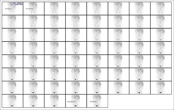
Tc-99m labeled red blood cell (RBC) scintigraphy is commonly used in the evaluation of acute gastrointestinal (GI) bleeding. On Tc-99m RBC studies, GI bleeding is seen as an initial focus of increased radiotracer activity that on subsequent images increases in intensity and changes position in a pattern that confirms to segments of bowel. We report a case of a patient with multiple episodes of GI bleeding referred to detect the source of bleeding. A Tc-99m labeled RBC scan was performed and the findings showed a focal abnormal hot spot in the mid quadrant of the abdomen, seen promptly in initial dynamic images. Subsequent static and single-photon emission computed tomography-CT (SPECT-CT) images found it to be umbilical varices. Most varices fill promptly as in this case and should not be misinterpreted as a focus of hemorrhage. SPECT-CT should be used in such cases so that that false-positive interpretation can be avoided.
Keywords: False-positive; gastrointestinal bleed scintigraphy; single-photon emission computed tomography-computed tomography; umbilical varices.
Conflict of interest statement
There are no conflicts of interest.
Figures


References
-
- Dam HQ, Brandon DC, Grantham VV, Hilson AJ, Howarth DM, Maurer AH, et al. The SNMMI procedure standard/EANM practice guideline for gastrointestinal bleeding scintigraphy 2.0. J Nucl Med Technol. 2014;42:308–17. - PubMed
-
- Howarth DM. The role of nuclear medicine in the detection of acute gastrointestinal bleeding. Semin Nucl Med. 2006;36:133–46. - PubMed
-
- Henkin RE, Bova D, Dillehay GL, Karesh SM, Halama JR, Wagner RH. Nuclear Medicine. 2nd ed. Philadelphia, PA: Mosby Elsevier; 2006. pp. 988–93.
