. 2018 Jul;16(1):755-760.
doi: 10.3892/ol.2018.8707. Epub 2018 May 14.

Thin-wall cystic lung cancer: A study of 45 cases

Affiliations

Thin-wall cystic lung cancer: A study of 45 cases

Hui Deng et al. Oncol Lett. 2018 Jul.

Abstract

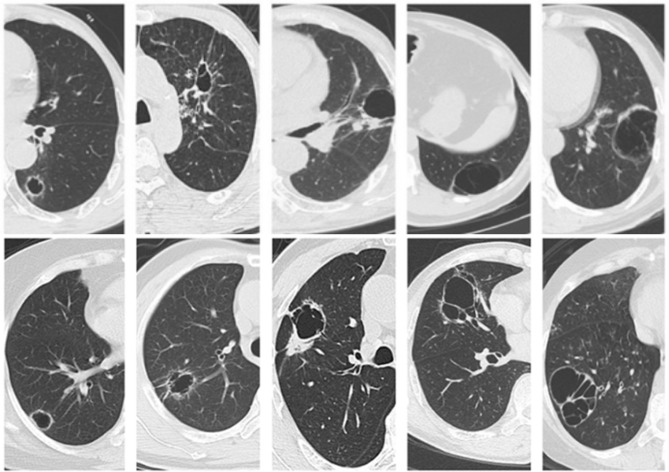
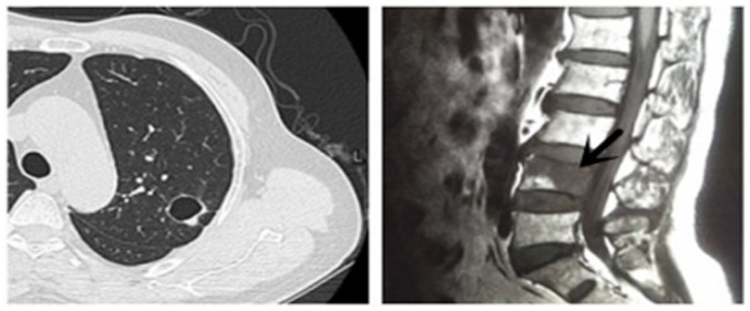
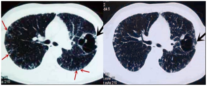
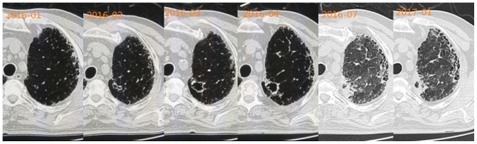
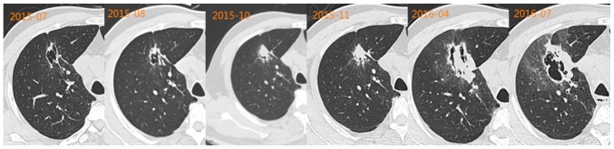
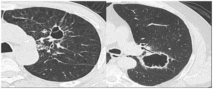
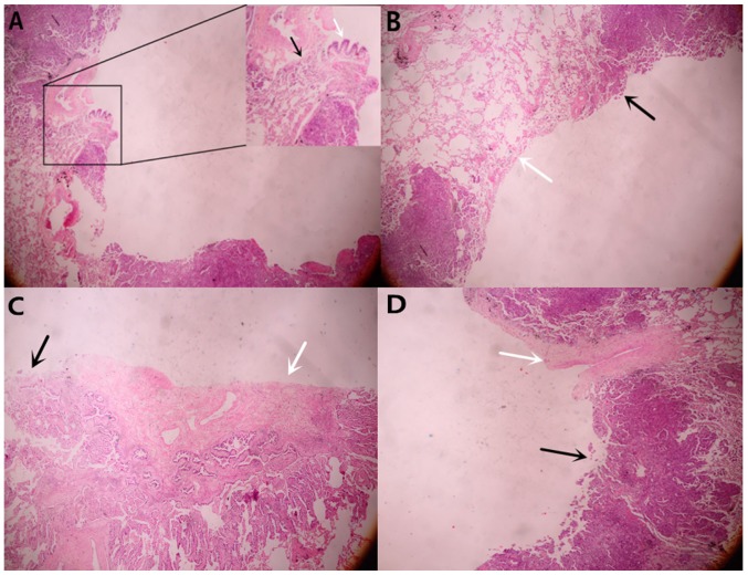
Thin-wall cystic lung cancer is uncommon. Consequently, there is a lack of knowledge concerning the features of this type of lung cancer, which may lead to misdiagnosis and delayed treatment. The aim of the present study is to understand the invasiveness and metastasis of thin-wall cystic lung cancer. The prognosis of this type of cancer will also be discussed. The present study attempted to determine the pathological interpretation of the imaging results. A total of 45 patients with this specific type of lung cancer were analyzed retrospectively based on the review of medical records, radiological findings, pathological changes and treatment strategies. Certain patients were also telephoned in order to learn about their recent physical conditions. Thin-wall cystic lung cancer displayed suspected malignant signs. The majority of these cases are adenocarcinoma, but certain cases of squamous cell carcinoma may also display cysts on their images. Although thin-wall cystic lung cancer is often thought to progress slowly, certain cases may progress rapidly. Distant metastasis, which is relatively rare, occurred in three cases. Cancer cells proliferate along the terminal bronchioles and destroy the lung tissues exposing the bronchial arteries and adjacent bronchi. Therefore, separation in cysts on the images was observed. In the majority of cases, the thin-wall cystic lung cancer proliferates slowly, but in a few cases it may be very aggressive.

Keywords: lung cancer; metastasis; pathology; prognosis; radiology.


Figures

Figure 1.
Figure 1.
Chest computed tomography presentations of solitary thin-wall cystic lung cancer.
Figure 2.
Figure 2.
Computed tomography image of thin-wall cystic lung cancer in a 57-year-old male (left). Metastases to the fourth lumbar spine (right).
Figure 3.
Figure 3.
A large, abnormal cystic lesion in the left lung of a 33-year-old male (black arrow) and multiple cystic lesions (red arrow) in the two lungs on computed tomography images.
Figure 4.
Figure 4.
The cystic wall of a 60-year-old male became irregularly thickened and certain malignant signs were observed over four consecutive months. The cyst is getting smaller and the parenchyma is increasing.
Figure 5.
Figure 5.
Adenocarcinoma with multiple lymph node metastases in a 51-year-old male. Upon follow-up examination the cyst is getting smaller and the parenchyma is increasing. A new cavity is eventually formed.
Figure 6.
Figure 6.
Two patients were diagnosed with squamous cell carcinoma by pathology. They also displayed cysts on the images.
Figure 7.
Figure 7.
(A) Tumor cells (black arrow) destroyed the bronchi wall (white arrow). (B) The cavity wall was covered with tumor cells in patients who underwent surgery. The black arrow represents the area of the wall covered with tumor cells and the white arrow represents the area of the wall not covered with tumor cells, which demonstrated that thin-wall cavity formation had been initiated prior to tumor cell covering the wall. (C) Hyperplastic fibrous tissue was observed inside the cyst. (D) The blood vessels (indicated by the white arrow) blocked the proliferation of tumor cells. Magnification, ×40.

References

    1. Chen Z, editor. Third national retrospect port-check of death-causation. 1st edition. Peking Union Medical College Press; Beijing, China: 2008. pp. 153–154.
    1. Ferlay J, Shin HR, Bray F, Forman D, Mathers C, Parkin DM. Estimates of worldwide burden of cancer in 2008: GLOBOCAN 2008. Int J Cancer. 2010;127:2893–2917. doi: 10.1002/ijc.25516. - DOI - PubMed
    1. Parkin DM, Bray F, Ferlay J, Pisani P. Global cancer statistics, 2002. CA Cancer J Clin. 2005;55:74–108. doi: 10.3322/canjclin.55.2.74. - DOI - PubMed
    1. Woodring JH. Pitfalls in the radiologic diagnosis of lung cancer. AJR Am J Roentgenol. 1990;154:1165–1175. doi: 10.2214/ajr.154.6.2110722. - DOI - PubMed
    1. Matsuoka T, Fukamitsu G, Onoda M, Uesugi N, Kawano K, Katou T. Synchronous multiple lung cancer including a lesion with a thin-wall cavity; report of a case. Kyobu Geka. 2010;63:164–167. (In Japanese) - PubMed

LinkOut - more resources