. 2018 Jun 15;9(46):28116-28130.
doi: 10.18632/oncotarget.25565.

Cancer stem cells from peritumoral tissue of glioblastoma multiforme: the possible missing link between tumor development and progression

Affiliations

Cancer stem cells from peritumoral tissue of glioblastoma multiforme: the possible missing link between tumor development and progression

Cristiana Angelucci et al. Oncotarget. .

Abstract

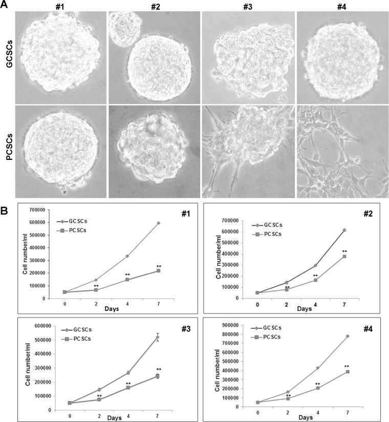
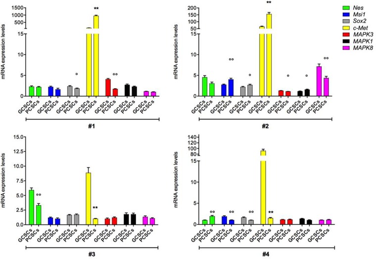
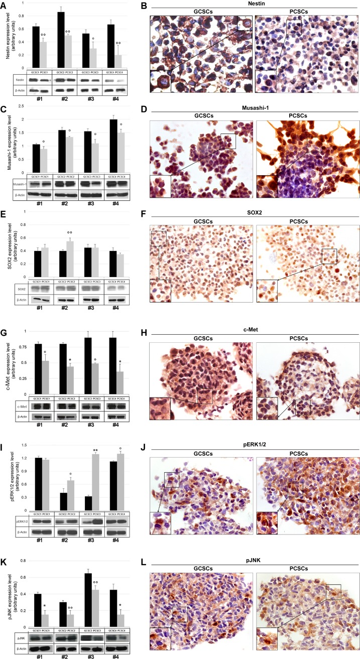
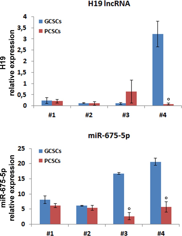
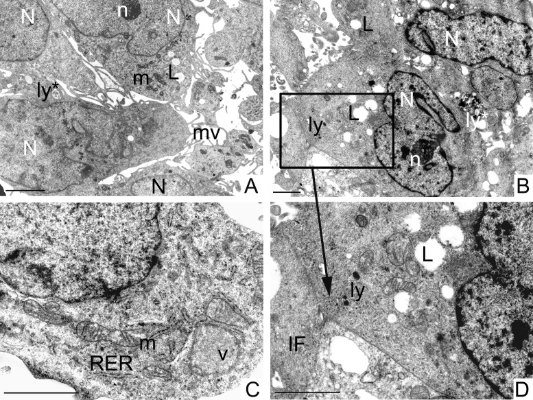
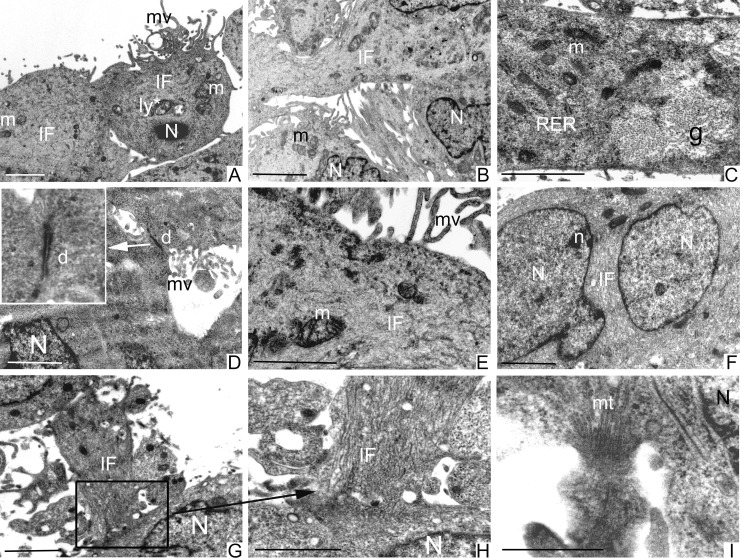
In glioblastoma multiforme (GBM), cancer stem cells (CSCs) are thought to be responsible for gliomagenesis, resistance to treatment and recurrence. Unfortunately, the prognosis for GBM remains poor and recurrence frequently occurs in the peritumoral tissue within 2 cm from the tumor edge. In this area, a population of CSCs has been demonstrated which may recapitulate the tumor after surgical resection. In the present study, we aimed to characterize CSCs derived from both peritumoral tissue (PCSCs) and GBM (GCSCs) in order to deepen their significance in GBM development and progression. The stemness of PCSC/GCSC pairs obtained from four human GBM surgical specimens was investigated by comparing the expression of specific stem cell markers such as Nestin, Musashi-1 and SOX2. In addition, the growth rate, the ultrastructural features and the expression of other molecules such as c-Met, pMet and MAP kinases, involved in cell migration/invasion, maintenance of tumor stemness and/or resistance to treatments were evaluated. Since it has been recently demonstrated the involvement of the long non-coding RNAs (lncRNAs) in the progression of gliomas, the expression of H19 lncRNA, as well as of one of its two mature products miR-675-5p was evaluated in neurospheres. Our results show significant differences between GCSCs and PCSCs in terms of proliferation, ultrastructural peculiarities and, at a lower extent, stemness profile. These differences might be important in view of their potential role as a therapeutic target.

Keywords: H19 lncRNA and miR-675-5p; glioblastoma cancer stem cells; peritumoral cancer stem cells; proliferation and invasiveness markers; stemness markers.


Conflict of interest statement

CONFLICTS OF INTEREST The authors declare no conflicts of interest.

Figures

Figure 1
Figure 1. Morphological and proliferation analysis of GCSC/PCSC pairs
(A) GCSCs derived from all the four patients, as well as PCSCs obtained from patients #1 and #2, grew as floating neurospheres. PCSCs corresponding to patients #3 and #4 grew as semi-adherent cells. Original magnification, ×400. (B) In each GCSC/PCSC pair (#1–4) analyzed, GCSCs (rumble) show a higher proliferation rate if compared to PCSCs (square). Values represent the mean ± SD of three independent experiments. Data were analyzed by Student t test, **p < 0.001 vs GCSCs.
Figure 2
Figure 2. Nes, Msi1, Sox2, c-Met, MAPK3, MAPK1 and MAPK8 gene expression in GCSC/PCSC pairs
The expression level of the indicated genes was evaluated by qPCR in GCSCs and PCSCs. The relative RNA quantity was normalized to GAPDH endogenous control. Nes (gene coding for Nestin protein), Msi1 (gene coding for Musashi-1 protein), Sox2 (gene coding for SOX2 protein), c-Met (gene coding for c-Met protein), MAPK3 (gene coding for ERK1 protein), MAPK1 (gene coding for ERK2 protein) and MAPK8 (gene coding for JNK protein). Bar graph show mean ± SE from three independent experiments. Data were analyzed by t-test pooled variance, °p < 0.05, °°p < 0.01 and **p < 0.001 vs GCSCs.
Figure 3
Figure 3. Nestin, Musashi-1, SOX2, c-Met, pERK1/2 and pJNK protein expression in GCSC/PCSC pairs
Nestin (A), Musashi-1 (C), SOX2 (E), c-Met (G), pERK1/2 (I) and pJNK (K) protein expression was evaluated by Western blot analysis. Representative blots from three separate experiments yielding similar results are shown. The intensity of bands was quantified by densitometric analysis and normalized to those of β-actin (used as loading control). The values shown represent the mean ± SD of three independent experiments. Data were analyzed by Student t-test, °p < 0.05, °°p < 0.01, *p < 0.005 and **p < 0.001 vs GCSCs. Immunocytochemical analysis of Nestin (B), Musashi-1 (D) and SOX2 (F), c-Met (H), pERK1/2 (J) and pJNK (L) performed on GCSC and PCSC frozen sections (10 μm). All the investigated markers were expressed in both cell populations. Nestin immunoreactivity was found at the level of cell cytoplasm (B). Musashi-1 specific staining was seen in both the cytoplasm and the nucleus (D). SOX2 staining was mainly detected in the nucleus (F). c-Met (H), pERK1/2 (J) and pJNK (L) immunoreactivity was present at the level of both the cytoplasm and the nucleus. Original magnification, ×630. Details of immuno-positive and -negative cells are shown at higher magnification in the bottom left insets (B, D, F, H, J, L).
Figure 4
Figure 4. H19 lncRNA and miR-675-5p expression in GCSC/PCSC pairs
The expression level of H19 lncRNA and miR-675-5p was evaluated by qPCR in GCSCs and PCSCs. All values were expressed as compared to those found in human astrocytes, where H19 expression was set = 1. Expression levels were normalized against U6 snRNA and TBP mRNA as controls for miR-675-5p or H19 lncRNA, respectively. The values shown represent the mean ± SD of two independent experiments. Data were analyzed by Student t-test; °p < 0.05 vs GCSCs.
Figure 5
Figure 5. Ultrastructural analysis of GCSCs
GCSCs showed irregular nuclei (N) mainly formed by euchromatin with marginated heterochromatin and prominent nucleoli (n) (A, B). Rough endoplasmic reticulum (RER), mitochondria (m), vesicles (v) and intermediate filaments (IF) were visible in the cytoplasm (C, D). Lipid droplets (L), primary and secondary lysosomes (ly and ly*, respectively) were observed (A, B and D). Plasma membranes showed microvilli (mv) (A) and loose cell-cell contacts (square in B and at higher magnification in D). Scale bars = 2 μm.
Figure 6
Figure 6. Ultrastructural analysis of PCSCs
PCSCs showed irregular nuclei (N) (A, B and F) and sometimes multiple nuclei with evident nucleoli (n) (F). Mitochondria (m) were visible (A–C, E). Cells were particularly rich in intermediate filaments (IF) located in the cytoplasm and in the perinuclear region (A, B, E–H). Microvilli (mv) were present on the cellular membrane (A, D and E). Abundant glycogen (g) stored free in the cytoplasm (C) was noted. Fully organized desmosomes (d) were also visible (D). Loose cell-cell contacts were established by cellular processes rich in IF (square in G and at higher magnification in H) and microtubules (mt) (I). Scale bars = 2 μm.

Similar articles

Cited by

References

    1. Magee JA, Piskounova E, Morrison SJ. Cancer stem cells: impact, heterogeneity, and uncertainty. Cancer Cell. 2012;21:283–296. - PMC - PubMed
    1. Fidoamore A, Cristiano L, Antonosante A, d’Angelo M, Di Giacomo E, Astarita C, Giordano A, Ippoliti R, Benedetti E, Cimini A. Glioblastoma stem cells microenvironment: the paracrine roles of the niche in drug and radioresistance. Stem Cells Int. 2016;2016:6809105. - PMC - PubMed
    1. Denysenko T, Gennero L, Roos MA, Melcarne A, Juenemann C, Faccani G, Morra I, Cavallo G, Reguzzi S, Pescarmona G, Ponzetto A. Glioblastoma cancer stem cells: heterogeneity, microenvironment and related therapeutic strategies. Cell Biochem Funct. 2010;28:343–351. - PubMed
    1. Bao S, Wu Q, McLendon RE, Hao Y, Shi Q, Hjelmeland AB, Dewhirst MW, Bigner DD, Rich JN. Glioma stem cells promote radioresistance by preferential activation of the DNA damage response. Nature. 2006;444:756–760. - PubMed
    1. Eramo A, Ricci-Vitiani L, Zeuner A, Pallini R, Lotti F, Sette G, Pilozzi E, Larocca LM, Peschle C, De Maria R. Chemotherapy resistance of glioblastoma stem cells. Cell Death Differ. 2006;13:1238–1241. - PubMed

LinkOut - more resources