Portal vein thrombosis: What surgeons need to know
- PMID: 29963409
- PMCID: PMC6018256
- DOI: 10.4103/IJCIIS.IJCIIS_71_17
Portal vein thrombosis: What surgeons need to know
Abstract
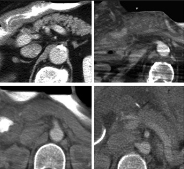
Key points: (a) The lifetime risk of portal vein thrombosis (PVT) is approximately 1%; (b) The portal vein is formed by the union of the splenic and superior mesenteric veins posterior to the pancreas; (c) Imaging modalities most frequently used to diagnose PVT include sonography, computed tomography, and magnetic resonance imaging; (d) Malignancy, hepatic cirrhosis, surgical trauma, and hypercoagulable conditions are the most common risk factors for the development of PVT; (e) PVT eventually leads to the formation of numerous collateral vessels around the thrombosed portal vein; (f) First-line treatment for PVT is therapeutic anticoagulation-it helps prevent the progression of the thrombotic process; (g) Other therapeutic options include surgery and interventional radiographic procedures including mechanical thrombectomy and thrombolysis; (h) Portal biliopathy is a clinicopathologic entity characterized by biliary abnormalities due to portal hypertension secondary to PVT and appears to be more common in cases of extrahepatic PVT.
Republished with permission from: Quarrie R, Stawicki SP. Portal vein thrombosis: What surgeons need to know. OPUS 12 Scientist 2008;2(3):30-33.
Keywords: Complications; pathophysiology; portal vein thrombosis; risk factors; therapeutic interventions.
Conflict of interest statement
There are no conflicts of interest.
Figures
References
-
- Condat B, Pessione F, Helene Denninger M, Hillaire S, Valla D. Recent portal or mesenteric venous thrombosis: Increased recognition and frequent recanalization on anticoagulant therapy. Hepatology. 2000;32:466–70. - PubMed
-
- Khan AN, MacDonald S, Sheen AJ, Sherlock D, Al-Khattab Y. Portal Vein Thrombosis. [Last accessed on 2008 Jun 10]. Available from: http://www.emedicine.com/radio/topic571.htm .
-
- Ricci P, Cantisani V, Biancari F, Drud FM, Coniglio M, Di Filippo A, et al. Contrast-enhanced color Doppler US in malignant portal vein thrombosis. Acta Radiol. 2000;41:470–3. - PubMed