Case Reports
. 2018 Jun;48(2):131-137.
doi: 10.5624/isd.2018.48.2.131. Epub 2018 Jun 19.

Recurrent symptomatic cemento-osseous dysplasia: A case report

Affiliations
Case Reports

Recurrent symptomatic cemento-osseous dysplasia: A case report

Chang-Ki Min et al. Imaging Sci Dent. 2018 Jun.

Abstract

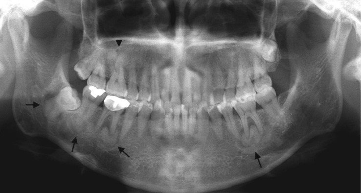
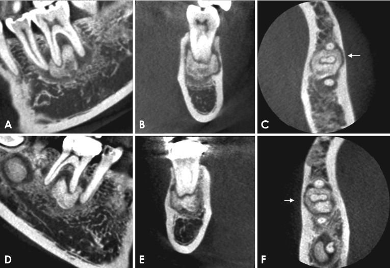
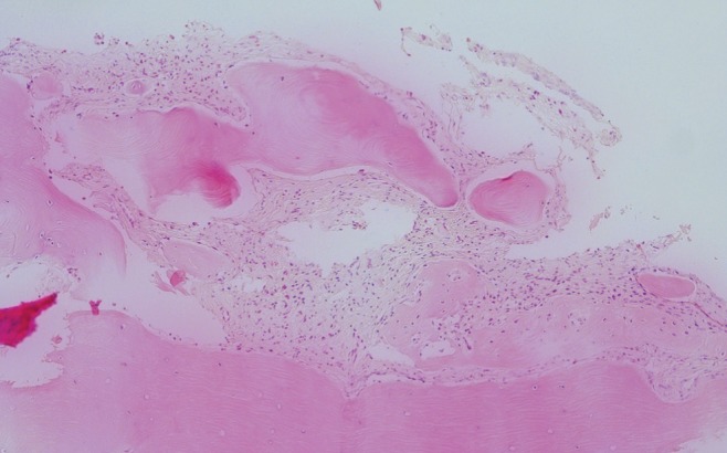
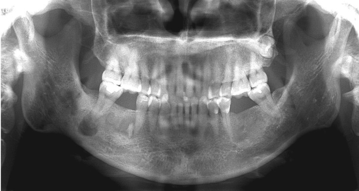
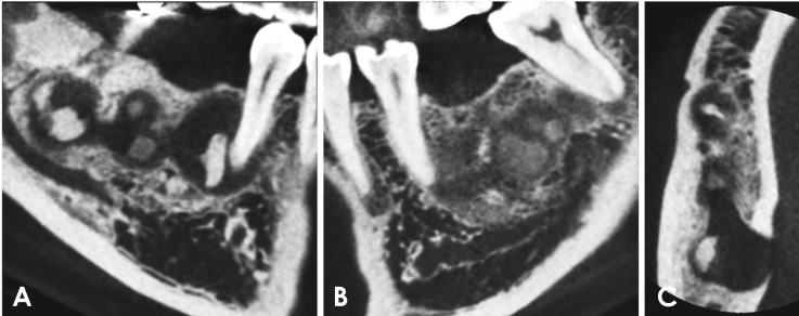
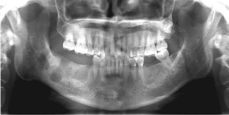
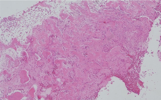
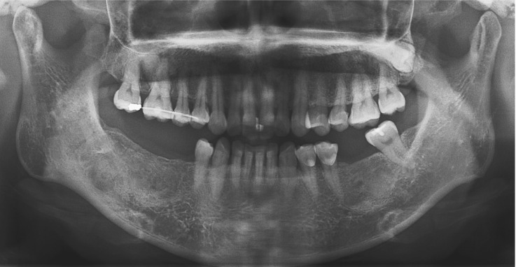
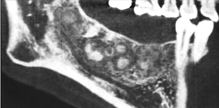
Cemento-osseous dysplasia (COD) is a benign fibro-osseous lesion of bone, in which normal bone is replaced by fibrous tissue, followed by calcification with osseous and cementum-like tissue. COD is classified into 3 categories according to its location: periapical, focal, and florid COD (FCOD). On radiography, FCOD appears radiolucent in its early stages. As it matures, radiopacities appear within the lesion, causing them to show a mixed appearance of radiolucency and radiopacity. Because FCOD is usually asymptomatic and grows in a self-limited manner, it does not require treatment. Secondary infection is the most frequent cause of symptomatic cases. We report a case of FCOD with symptoms that appeared after a dental restoration procedure and persisted after repeated operations. The purpose of this report is to emphasize the importance of thorough radiological evaluations of patients with FCOD before treatment.

Keywords: Bone Diseases; Cone-Beam Computed Tomography; Florid Cemento-osseous Dysplasia; Radiography, Panoramic.


Figures

Fig. 1
Fig. 1. Panoramic image shows multiple sclerotic masses with radiolucent rims in the apical region of the left mandibular second molar and the right mandibular first, second, and third molars (arrows), as well as completely radiopaque masses in the apical region of the right maxillary first molar and the left maxillary second premolar (arrowhead).
Fig. 2
Fig. 2. Cone-beam computed tomography scans reveal well-defined radiopaque masses surrounded by radiolucent rims in the periapical regions of the left mandibular second premolar and first molar (A–C) and the right mandibular first and second molar (D–F). All the lesions were separated from the adjacent teeth by a radiolucent line. Invasion of the cortical bone can be observed (arrows). (A and D: sagittal, B and E: frontal, C and F: axial scan)
Fig. 3
Fig. 3. Scintigraphs show increased radiopharmaceutical uptake in the posterior maxilla and mandible.
Fig. 4
Fig. 4. Histopathologic examination shows multiple areas of irregular woven bone in dense fibrous stroma without any fibrous capsules, representing cemento-osseous dysplasia (H&E stain, original magnification ×100).
Fig. 5
Fig. 5. Panoramic image 3 years after surgery shows the remaining calcified masses around the right mandibular first molar and increased periapical radiolucency at the right mandibular second molar.
Fig. 6
Fig. 6. A and B. Cone-beam computed tomography (CBCT) scans 3 years after surgery reveal the remaining and newly-formed radiopacities inside the lesion. C. Axial CBCT scan shows perforation of the lingual cortical plate near the right mandibular second molar.
Fig. 7
Fig. 7. Panoramic image obtained immediately after secondary surgery shows that all the lesions of the right mandible had been removed.
Fig. 8
Fig. 8. Histopathologic finding shows numerous areas of woven bone in fibrous connective tissue with infiltration of inflammatory cells (H&E stain, original magnification ×40).
Fig. 9
Fig. 9. Panoramic image demonstrates sclerotic masses surrounded by radiolucent halos in the right and left mandible at the previous surgical sites.
Fig. 10
Fig. 10. Sagittal cone-beam computed tomography scan clearly reveals sclerotic masses surrounded by radiolucent halos in the right mandible at the previous surgical sites.

Similar articles

Cited by

References

    1. MacDonald DS. Maxillofacial fibro-osseous lesions. Clin Radiol. 2015;70:25–36. - PubMed
    1. MacDonald-Jankowski DS. Florid cemento-osseous dysplasia: a systematic review. Dentomaxillofac Radiol. 2003;32:141–149. - PubMed
    1. Mainville GN, Turgeon DP, Kauzman A. Diagnosis and management of benign fibro-osseous lesions of the jaws: a current review for the dental clinician. Oral Dis. 2017;23:440–450. - PubMed
    1. Su L, Weathers DR, Waldron CA. Distinguishing features of focal cemento-osseous dysplasia and cemento-ossifying fibromas. II. A clinical and radiologic spectrum of 316 cases. Oral Surg Oral Med Oral Pathol Oral Radiol Endod. 1997;84:540–549. - PubMed
    1. Cavalcanti PH, Nascimento EH, Pontual ML, Pontual AD, Marcelos PG, Perez DE, et al. Cemento-osseous dysplasias: imaging features based on cone beam computed tomography scans. Braz Dent J. 2018;29:99–104. - PubMed

Publication types