. 2018 Oct;17(5):e12800.
doi: 10.1111/acel.12800. Epub 2018 Jul 2.

FOXO are required for intervertebral disk homeostasis during aging and their deficiency promotes disk degeneration

Affiliations

FOXO are required for intervertebral disk homeostasis during aging and their deficiency promotes disk degeneration

Oscar Alvarez-Garcia et al. Aging Cell. 2018 Oct.

Abstract

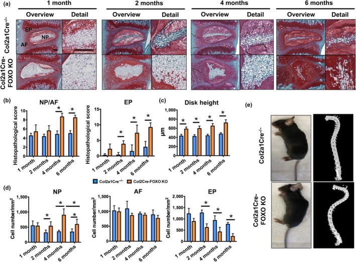
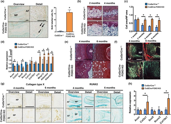
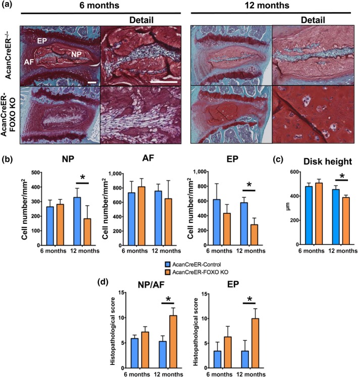
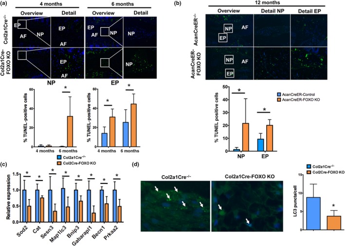
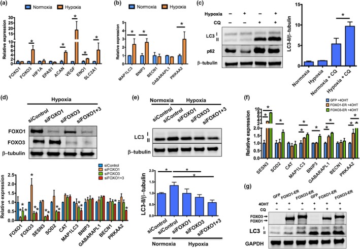
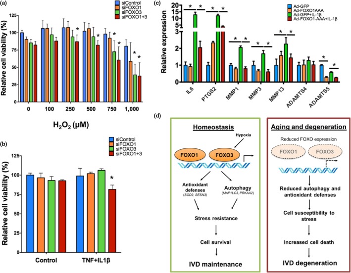
Intervertebral disk (IVD) degeneration is a prevalent age-associated musculoskeletal disorder and a major cause of chronic low back pain. Aging is the main risk factor for the disease, but the molecular mechanisms regulating IVD homeostasis during aging are unknown. The aim of this study was to investigate the function of FOXO, a family of transcription factors linked to aging and longevity, in IVD aging and age-related degeneration. Conditional deletion of all FOXO isoforms (FOXO1, 3, and 4) in IVD using the Col2a1Cre and AcanCreER mouse resulted in spontaneous development of IVD degeneration that was driven by severe cell loss in the nucleus pulposus (NP) and cartilaginous endplates (EP). Conditional deletion of individual FOXO in mature mice showed that FOXO1 and FOXO3 are the dominant isoforms and have redundant functions in promoting IVD homeostasis. Gene expression analyses indicated impaired autophagy and reduced antioxidant defenses in the NP of FOXO-deficient IVD. In primary human NP cells, FOXO directly regulated autophagy and adaptation to hypoxia and promoted resistance to oxidative and inflammatory stress. Our findings demonstrate that FOXO are critical regulators of IVD homeostasis during aging and suggest that maintaining or restoring FOXO expression can be a therapeutic strategy to promote healthy IVD aging and delay the onset of IVD degeneration.

Keywords: FOXO; aging; autophagy; intervertebral disk.


Figures

Figure 1
Figure 1
Impaired intervertebral disk maturation and spontaneous degeneration in mice with conditional deletion of FOXO. (a) Safranin O staining in lumbar intervertebral disk (IVD) samples isolated from Col2a1Cre−/− and Col2a1Cre‐FOXO KO mice at 1, 2, 4, and 6 months of age (n = 6–8 mice per group). NP: nucleus pulposus; AF: annulus fibrosus; EP: endplate. Magnification bar = 100 µm. (b) Histopathological scores in the nucleus NP/AF and EP of lumbar IVD samples isolated from Col2a1Cre−/− and Col2a1Cre‐FOXO KO mice at 1, 2, 4, and 6 months of age (n = 6–8 mice per group). (c) Measurement of disk height of lumbar IVD samples from Col2a1Cre−/− and Col2a1Cre‐FOXO KO mice at 1, 2, 4, and 6 months of age (n = 6–8 mice per group). (d) Quantification of cellularity in the NP, AF, and EP of lumbar IVD samples isolated from Col2a1Cre−/− and Col2a1Cre‐FOXO KO at 1, 2, 4, and 6 months of age (n = 6–8 mice per group). (e) Photograph (left) and µ‐CT image (right) of 6‐month‐old Col2a1Cre−/− and Col2a1Cre‐FOXO TKO mice. Values shown are mean ± SD. Statistical comparisons were assessed by one‐way analysis of variance (ANOVA) followed by a post hoc Tukey's test. *p < 0.05
Figure 2
Figure 2
Regulation of intervertebral disk proliferation and differentiation by FOXO. (a) Immunohistochemical detection of 5‐bromo‐2'‐deoxyuridine (BrdU) in lumbar intervertebral disk (IVD) samples from Col2a1Cre−/− and Col2a1Cre‐FOXO KO mice at 1 month of age (n = 3 mice per group) showing immunopositive cells (black arrows) in the nucleus pulposus (NP). AF: annulus fibrosus. Panel on the right shows quantification of BrdU‐positive cells in the NP. Magnification bar = 100 µm. (b) Safranin O staining in NP from lumbar IVD samples isolated from Col2a1Cre−/− and Col2a1Cre‐FOXO KO mice at 2 and 4 months of age (n = 6–8 mice per group). Magnification bar = 50 µm. (c) Histomorphometric quantification of the ratio of the NP cell area to total NP area in IVD from Col2a1Cre−/− and Col2a1Cre‐FOXO KO mice at 1, 2, 4, and 6 months of age (n = 6–8 mice per group). (d) Gene expression analysis of anabolic and catabolic genes in the NP of lumbar IVD samples from Col2a1Cre−/− and Col2a1Cre‐FOXO KO at 4 months of age (n = 4 mice per group). (e) Safranin O staining in annulus fibrosus (AF) of lumbar IVD isolated from Col2a1Cre−/− and Col2a1Cre‐FOXO KO mice at 4 and 6 months of age. Magnification bar = 100 µm. (f) Picrosirius red staining in lumbar IVD isolated from Col2a1Cre−/− and Col2a1Cre‐FOXO KO mice at 4 and 6 months of age (n = 6 mice per group). Images were obtained under polarized light to show collagen birefringence. Magnification bar = 100 µm. (g) Collagen type X (left panel) and RUNX2 (right panel) immunohistochemistry in lumbar IVD from Col2a1Cre−/− and Col2a1Cre‐FOXO KO mice at 4 and 6 months of age (n = 5 mice per group). Magnification bar = 100 µm. (h) Gene expression analysis in the AF of lumbar IVD samples from Col2a1Cre−/− and Col2a1Cre‐FOXO KO at 4 months of age (n = 4 mice per group). Values shown are mean ± SD. Statistical comparisons were assessed by an unpaired, two‐tailed t‐test after testing for equal variance using an F‐test. Values are mean ± SD. *p < 0.05
Figure 3
Figure 3
Spontaneous intervertebral disk degeneration in skeletally mature mice with conditional deletion of FOXO. (a) Safranin O staining in lumbar intervertebral disk (IVD) from AcanCreER−/− and AcanCreER‐FOXO KO mice at 6 and 12 months of age (n = 6–8 mice per group). NP: nucleus pulposus; AF: annulus fibrosus; EP: endplate. Magnification bar = 100 µm. (b) Quantification of cellularity in NP, AF, and EP of lumbar IVD from AcanCreER−/− and AcanCreER‐FOXO KO mice at 6 and 12 months of age (n = 6–8 mice per group). (c) Measurement of disk height of lumbar IVD samples AcanCreER−/− and AcanCreER‐FOXO KO mice at 6 and 12 months of age (n = 6–8 mice per group). (d) Histopathological scores in the NP/AF and EP of lumbar IVD from AcanCreER−/− and AcanCreER‐FOXO KO mice at 6 and 12 months of age (n = 6–8 mice per group). Values shown are mean ± SD. Statistical comparisons were assessed by an unpaired, two‐tailed t‐test after testing for equal variance using an F‐test. Values are mean ± SD. *p < 0.05
Figure 4
Figure 4
Reduced nucleus pulposus cell viability and impaired autophagy in FOXO‐deficient intervertebral disks. (a) Terminal deoxynucleotidyl transferase (TdT) dUTP Nick‐End Labeling (TUNEL) staining in lumbar intervertebral disks (IVD) isolated from Col2a1Cre−/− and Col2a1Cre‐FOXO KO mice at 4 and 6 months of age (n = 5 mice per group). Lower panels show quantification of TUNEL‐positive cells in the NP and EP. No positive cells were observed in the AF. NP: nucleus pulposus; AF: annulus fibrosus; EP: endplate. Magnification bar = 100 µm. (b) TUNEL staining in lumbar IVD from AcanCreER−/− and AcanCreER‐FOXO KO mice at 12 months of age (n = 5 mice per group). Lower panels show quantification of TUNEL‐positive cells in the NP and EP. (c) Gene expression analysis of homeostatic genes in NP from Col2a1Cre−/− and Col2a1Cre‐FOXO KO mice at 2 months of age (n = 4 mice per group). (d) Immunofluorescence staining for LC3 in the NP of lumbar IVD isolated from Col2a1Cre−/− and Col2a1Cre‐FOXO KO mice at 4 months of age (n = 5 mice per group) shows a decrease in immunostained cells in FOXO‐deficient NP cells. Right panel shows quantification of LC3 puncta per cell. Values shown are mean ± SD. Statistical comparisons were assessed by an unpaired, two‐tailed t‐test after testing for equal variance using an F‐test. Values are mean ± SD. *p < 0.05
Figure 5
Figure 5
FOXO mediate hypoxia‐induced autophagy in human nucleus pulposus cells. (a,b) Gene expression analysis in human primary nucleus pulposus (NP) cells cultured under normoxia (20% oxygen) or hypoxia (1% oxygen) for 24 hr showing hypoxia‐induced activation of FOXO3 and HIF1A signaling (a) and autophagic genes (b). (c) Western blot analysis of LC3‐I, LC3‐II, and p62 protein levels in human NP cells cultured in normoxia or hypoxia for 24 hr in the presence or absence of 25 µM chloroquine (CQ). Panel on the right shows densitometric quantification of LC3‐II and β‐tubulin. Values shown are mean ± SD of three different experiments. (d) Human NP cells were transfected with siRNA specific for FOXO1 (siFOXO1), FOXO3 (siFOXO3), or a combination of both (siFOXO1 + 3) and cultured in hypoxia for 24 hr. Upper panel shows Western blot analysis of FOXO1 and FOXO3 proteins confirming FOXO knockdown. Lower panel shows gene expression analysis of antioxidant and autophagic genes upon FOXO knockdown. (e) Western blot analysis of LC3 protein levels in human NP cells transfected with the indicated siRNA and cultured in normoxia or hypoxia for 24 hr in the presence of 25 µM CQ. Lower panel shows densitometric quantification of LC3‐II and β‐tubulin. (f) Gene expression analysis in human immortalized NP cells transfected with plasmids encoding for GFP, FOXO1‐ER, or FOXO3‐ER and treated with 1 µM 4OHT for 24 hr. (g) Western blot analysis of FOXO1, FOXO3, and LC3 protein levels in human NP cells transfected with plasmids encoding for GFP, FOXO1‐ER, or FOXO3‐ER and treated with 1 µM 4‐hydroxytamoxifen (4OHT) for 24 hr in the presence or absence of 25 µM CQ. Values shown are mean ± SD. Statistical comparisons were assessed by an unpaired, two‐tailed t‐test after testing for equal variance using an F‐test. *p < 0.05
Figure 6
Figure 6
FOXO promote resistance to oxidative and inflammatory stress in human nucleus pulposus cells. (a, b) Cell viability analysis in human NP cells transfected with siRNA specific for FOXO1 (siFOXO1), FOXO3 (siFOXO3), or a combination of both (siFOXO1 + 3) and treated with different H2O2 concentrations (a) or TNF (10 ng/ml) and IL‐1β (10 ng/ml) (b) for 24 hr. (c) Cells from normal human NP were transduced with adenovirus encoding green fluorescent protein (GFP) or a constitutively active FOXO1 mutant (FOXO1‐AAA) and stimulated with IL‐1β (1 ng/ml). RNA was isolated after 6 hr for qPCR analysis. Values shown are mean ± SD of three different experiments performed in duplicate. Statistical comparisons were assessed by an unpaired, two‐tailed t‐test after testing for equal variance using an F‐test. *p < 0.05. (d) Schematic representation of FOXO mechanisms in regulating intervertebral disk (IVD) maturation and homeostasis. NP: nucleus pulposus; AF: annulus fibrosus; EP: endplate

References

    1. Akasaki, Y. , Alvarez‐Garcia, O. , Saito, M. , Carames, B. , Iwamoto, Y. , & Lotz, M. K. (2014). FoxO transcription factors support oxidative stress resistance in human chondrocytes. Arthritis & Rheumatology, 66, 3349–3358. 10.1002/art.38868 - DOI - PMC - PubMed
    1. Alvarez‐Garcia, O. , Matsuzaki, T. , Olmer, M. , Masuda, K. , & Lotz, M. K. (2017). Age‐related reduction in the expression of FOXO transcription factors and correlations with intervertebral disc degeneration. Journal of Orthopaedic Research, 35, 2682–2691. - PMC - PubMed
    1. Alvarez‐Garcia, O. , Matsuzaki, T. , Olmer, M. , Plate, L. , Kelly, J. W. , & Lotz, M. K. (2017). Regulated in development and DNA damage response 1 deficiency impairs autophagy and mitochondrial biogenesis in articular cartilage and increases the severity of experimental osteoarthritis. Arthritis & Rheumatology, 69, 1418–1428. - PMC - PubMed
    1. Ambrogini, E. , Almeida, M. , Martin‐Millan, M. , Paik, J. H. , Depinho, R. A. , Han, L. , … Manolagas, S. C. (2010). FoxO‐mediated defense against oxidative stress in osteoblasts is indispensable for skeletal homeostasis in mice. Cell Metabolism, 11, 136–146. 10.1016/j.cmet.2009.12.009 - DOI - PMC - PubMed
    1. Anderson, D. G. , & Tannoury, C. (2005). Molecular pathogenic factors in symptomatic disc degeneration. The Spine Journal, 5, 260S–266S. - PubMed

Publication types