

Vitamin D Regulation of the Uridine Phosphorylase 1 Gene and Uridine-Induced DNA Damage in Colon in African Americans and European Americans
- PMID: 29964038
- PMCID: PMC6866230
- DOI: 10.1053/j.gastro.2018.06.049
Vitamin D Regulation of the Uridine Phosphorylase 1 Gene and Uridine-Induced DNA Damage in Colon in African Americans and European Americans
Abstract
Background & aims: African Americans have the greatest colorectal cancer (CRC) burden in the United States; interethnic differences in protective effects of vitamin D might contribute to disparities. 1α,25(OH)2D3 vitamin D (the active form of vitamin D) induces transcription of the uridine phosphorylase gene (UPP1) in colon tissues of European Americans but to a lesser extent in colon tissues of African Americans. UPP1-knockout mice have increased intestinal concentrations of uridine and Deoxyuridine triphosphate (dUTP), have increased uridine-induced DNA damage, and develop colon tumors. We studied 1α,25(OH)2D3 regulation of UPP1 and uridine-induced DNA damage in the colon and differences in these processes between African and European Americans.
Methods: We quantified expression and activity of UPP1 in response to 1α,25(OH)2D3 in young adult mouse colonic cells, human CRC cells (LS174T), and organoids (derived from rectosigmoid biopsy samples of healthy individuals undergoing colonoscopies) using quantitative polymerase chain reaction, immunoblot, and immunocytochemistry assays. Binding of the vitamin D receptor to UPP1 was tested by chromatin immunoprecipitation. Uridine-induced DNA damage was measured by fragment-length analysis in repair enzyme assays. Allele-specific 1α,25(OH)2D3 responses were tested using luciferase assays.
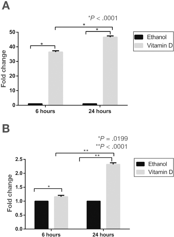
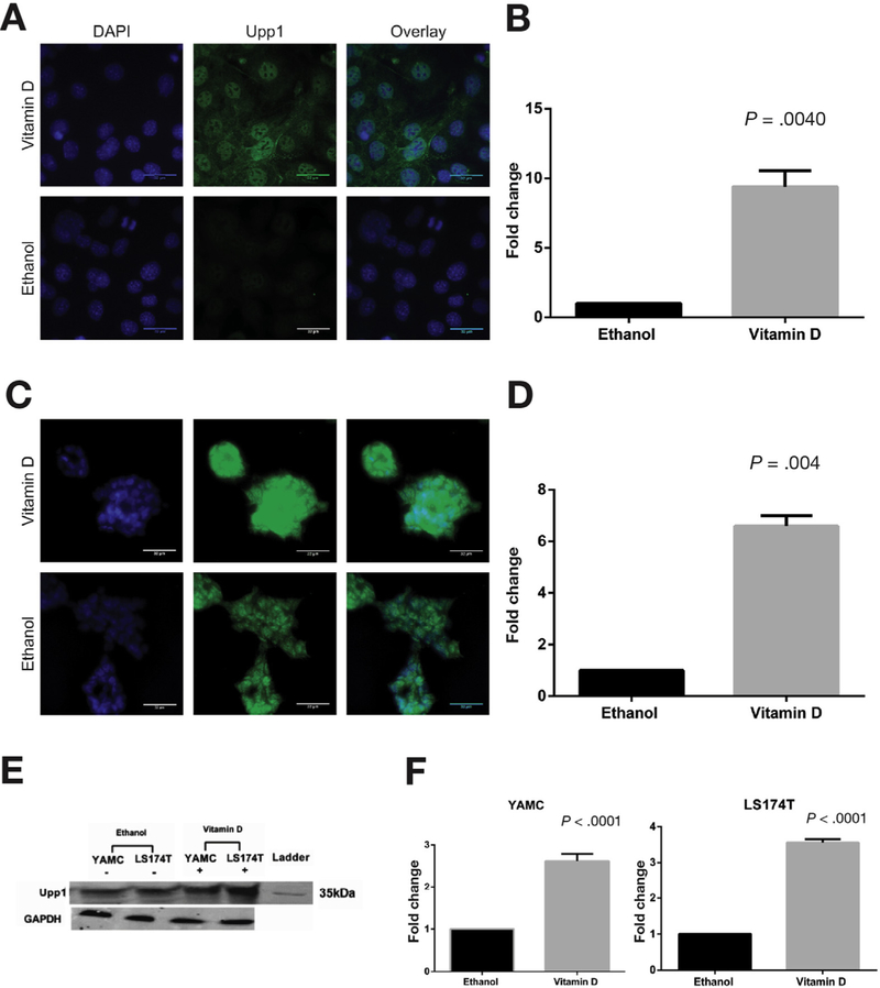
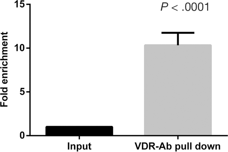
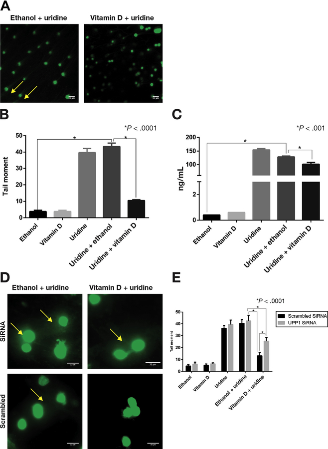
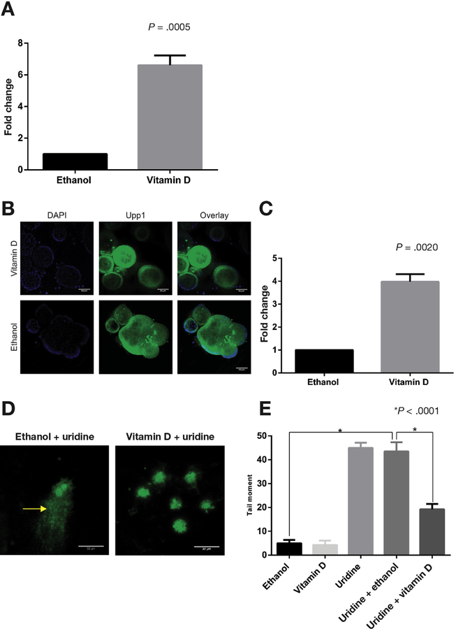
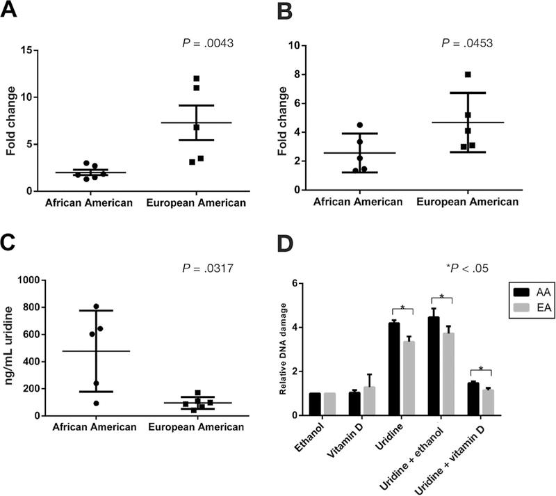
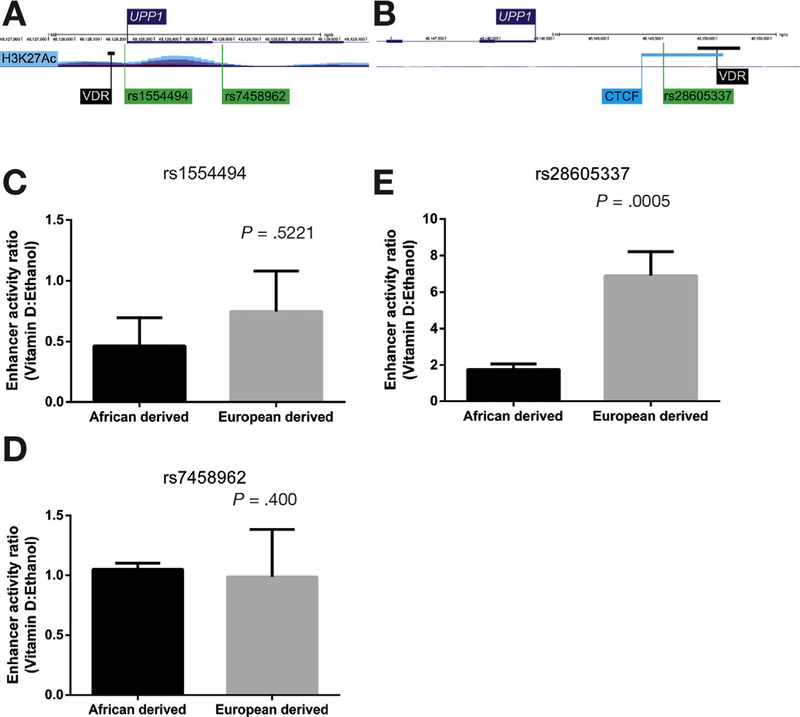
Results: Vitamin D increased levels of UPP1 mRNA, protein, and enzymatic activity and increased vitamin D receptor binding to the UPP1 promoter in young adult mouse colonic cells, LS174T cells, and organoids. 1α,25(OH)2D3 significantly reduced levels of uridine and uridine-induced DNA damage in these cells, which required UPP1 expression. Organoids derived from colon tissues of African Americans expressed lower levels of UPP1 after exposure to 1α,25(OH)2D3 and had increased uridine-induced DNA damage compared with organoids derived from tissues of European Americans. Luciferase assays with the T allele of single nucleotide polymorphism rs28605337 near UPP1, which is found more frequently in African Americans than European Americans, expressed lower levels of UPP1 after exposure to 1α,25(OH)2D3 than assays without this variant.
Conclusions: We found vitamin D to increase expression of UPP1, leading to reduce uridine-induced DNA damage, in colon cells and organoids. A polymorphism in UPP1 found more frequently in African Americans than European Americans reduced UPP1 expression upon cell exposure to 1α,25(OH)2D3. Differences in expression of UPP1 in response to vitamin D could contribute to the increased risk of CRC in African Americans.
Keywords: CRC; Ethnicity; Genetics; Risk Factor.
Copyright © 2018 AGA Institute. Published by Elsevier Inc. All rights reserved.
Conflict of interest statement
Conflicts of interest
The authors disclose no conflicts.
Figures
Similar articles
-
Genomic and epigenomic active vitamin D responses in human colonic organoids.Physiol Genomics. 2021 Jun 1;53(6):235-248. doi: 10.1152/physiolgenomics.00150.2020. Epub 2021 Apr 26. Physiol Genomics. 2021. PMID: 33900108 Free PMC article.
-
Colonic transcriptional response to 1α,25(OH)2 vitamin D3 in African- and European-Americans.J Steroid Biochem Mol Biol. 2017 Apr;168:49-59. doi: 10.1016/j.jsbmb.2017.02.001. Epub 2017 Feb 3. J Steroid Biochem Mol Biol. 2017. PMID: 28163244 Free PMC article.
-
Vitamin D receptor activation down-regulates the small heterodimer partner and increases CYP7A1 to lower cholesterol.Gastroenterology. 2014 Apr;146(4):1048-59. doi: 10.1053/j.gastro.2013.12.027. Epub 2013 Dec 21. Gastroenterology. 2014. PMID: 24365583
-
1alpha(OH)D3 One-alpha-hydroxy-cholecalciferol--an active vitamin D analog. Clinical studies on prophylaxis and treatment of secondary hyperparathyroidism in uremic patients on chronic dialysis.Dan Med Bull. 2008 Nov;55(4):186-210. Dan Med Bull. 2008. PMID: 19232159 Review.
-
Differential response to 1α,25-dihydroxyvitamin D3 (1α,25(OH)2D3) in non-small cell lung cancer cells with distinct oncogene mutations.J Steroid Biochem Mol Biol. 2013 Jul;136:264-70. doi: 10.1016/j.jsbmb.2012.09.022. Epub 2012 Sep 28. J Steroid Biochem Mol Biol. 2013. PMID: 23026510 Free PMC article. Review.
Cited by
-
Vitamin D supplementation and colorectal cancer prognosis.Med Oncol. 2019 Jun 12;36(8):69. doi: 10.1007/s12032-019-1293-x. Med Oncol. 2019. PMID: 31190098 No abstract available.
-
Genomic and epigenomic responses to aspirin in human colonic organoids.Physiol Genomics. 2023 Mar 1;55(3):101-112. doi: 10.1152/physiolgenomics.00070.2022. Epub 2023 Jan 16. Physiol Genomics. 2023. PMID: 36645669 Free PMC article.
-
VDR Signaling via the Enzyme NAT2 Inhibits Colorectal Cancer Progression.Front Pharmacol. 2021 Nov 16;12:727704. doi: 10.3389/fphar.2021.727704. eCollection 2021. Front Pharmacol. 2021. PMID: 34867333 Free PMC article.
-
Genomic and epigenomic active vitamin D responses in human colonic organoids.Physiol Genomics. 2021 Jun 1;53(6):235-248. doi: 10.1152/physiolgenomics.00150.2020. Epub 2021 Apr 26. Physiol Genomics. 2021. PMID: 33900108 Free PMC article.
-
UPP1 promotes lung adenocarcinoma progression through the induction of an immunosuppressive microenvironment.Nat Commun. 2024 Feb 8;15(1):1200. doi: 10.1038/s41467-024-45340-w. Nat Commun. 2024. PMID: 38331898 Free PMC article.
References
-
- American Cancer Society. Colorectal cancer: preventable, beatable, treatable. Volume 2017, Feburary 2017.
-
- Myers RE, Ruth K, Manne SL, et al. Effects of genetic and environmental risk assessment feedback on colorectal cancer screening adherence. J Behav Med 2015;38: 777–786. - PubMed
-
- Akin H, Tozun N. Diet, microbiota, and colorectal cancer. J Clin Gastroenterol 2014;48(Suppl 1):S67–S69. - PubMed
-
- Giovannucci E. The epidemiology of vitamin D and cancer incidence and mortality: a review (United States). Cancer Causes Control 2005;16:83–95. - PubMed
Publication types
MeSH terms
Substances
Grants and funding
LinkOut - more resources
Full Text Sources
Other Literature Sources
