. 2018 Oct;147(2):178-189.
doi: 10.1111/jnc.14543. Epub 2018 Sep 10.

The combination of ribose and adenine promotes adenosine release and attenuates the intensity and frequency of epileptiform activity in hippocampal slices: Evidence for the rapid depletion of cellular ATP during electrographic seizures

Affiliations

The combination of ribose and adenine promotes adenosine release and attenuates the intensity and frequency of epileptiform activity in hippocampal slices: Evidence for the rapid depletion of cellular ATP during electrographic seizures

Jessicka Hall et al. J Neurochem. 2018 Oct.

Abstract

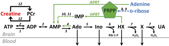
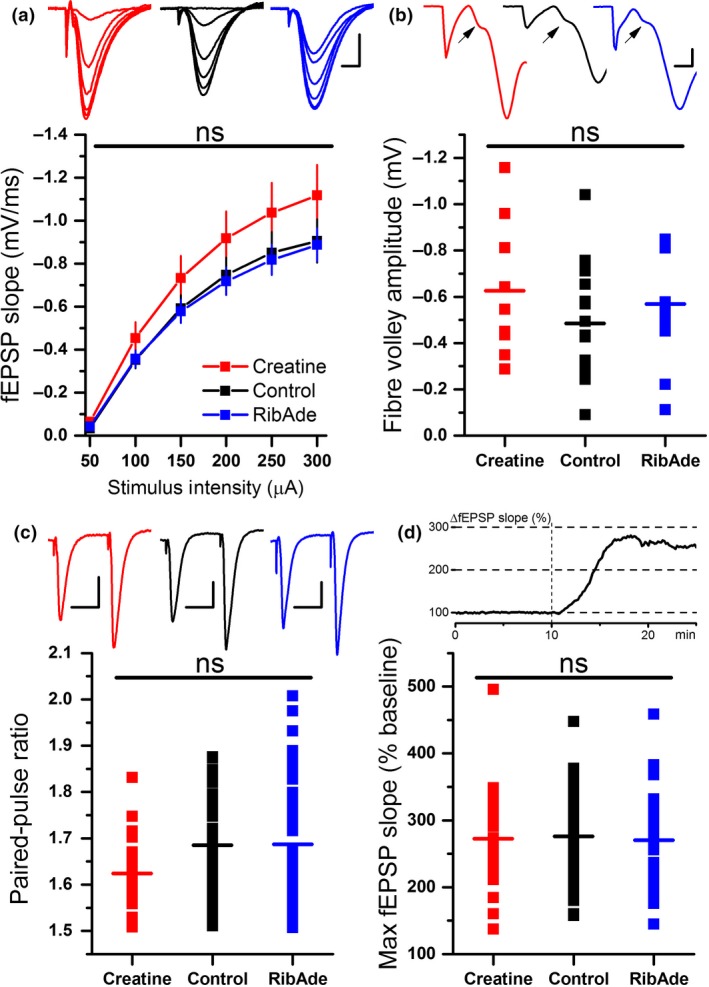
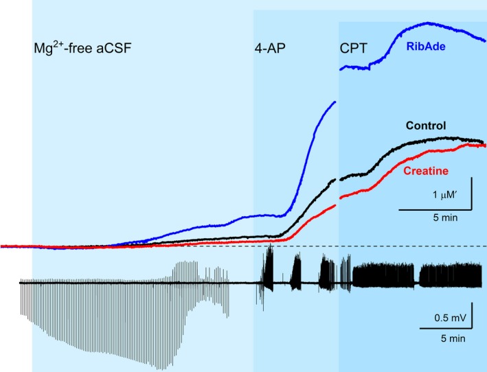
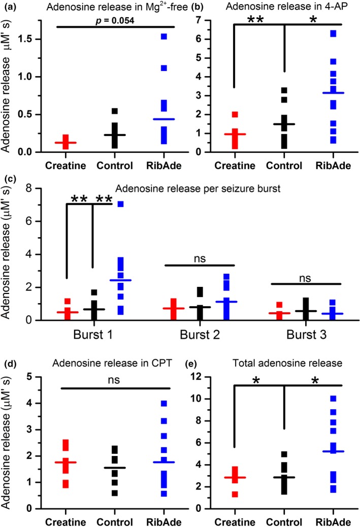
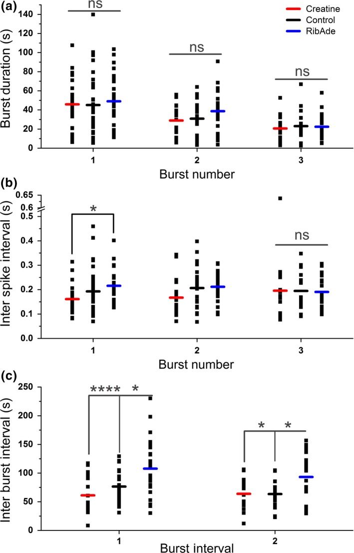
In addition to being the universal cellular energy source, ATP is the primary reservoir for the neuromodulator adenosine. Consequently, adenosine is produced during ATP-depleting conditions, such as epileptic seizures, during which adenosine acts as an anticonvulsant to terminate seizure activity and raise the threshold for subsequent seizures. These actions protect neurones from excessive ionic fluxes and hence preserve the remaining cellular content of ATP. We have investigated the consequences of manipulation of intracellular ATP levels on adenosine release and epileptiform activity in hippocampal slices by pre-incubating slices (3 h) with creatine (1 mM) and the combination of ribose (1 mM) and adenine (50 μM; RibAde). Creatine buffers and protects the concentration of cellular ATP, whereas RibAde restores the reduced cellular ATP in brain slices to near physiological levels. Using electrophysiological recordings and microelectrode biosensors for adenosine, we find that, while having no effect on basal synaptic transmission or paired-pulse facilitation, pre-incubation with creatine reduced adenosine release during Mg2+- free/4-aminopyridine-induced electrographic seizure activity, whereas RibAde increased adenosine release. This increased release of adenosine was associated with an attenuation of both the intensity and frequency of seizure activity. Given the depletion of ATP after injury to the brain, the propensity for seizures after trauma and the risk of epileptogenesis, therapeutic strategies elevating the cellular reservoir of adenosine may have value in the traumatized brain. Ribose and adenine are both in use in man and thus their combination merits consideration as a potential therapeutic for the acutely injured central nervous system.

Keywords: ATP; Adenosine; RibAde; epilepsy; purines; seizures.


Figures

Figure 1
Figure 1
ATP metabolism and synthesis via the cytosolic purine salvage pathway. Under conditions of energy depletion, ATP is metabolized to adenosine (Ado), inosine (Ino), hypoxanthine (HX) and xanthine (X), which can, via equilibrative transporters, leave cells and enter the bloodstream. Direct cellular release of ATP and subsequent extracellular metabolism by ectonucleotidases (not shown) provides another source of extracellular adenosine and purine loss to the bloodstream. Purine salvage (green arrows) restores adenine nucleotide levels via adenine phosphoribosyl‐transferase (APRT, EC 2.4.2.7; adenine to AMP) and hypoxanthine phosphoribosyl‐transferase (HPRT, EC 2.4.2.8; hypoxanthine to inosine monophosphate, IMP). This reaction requires 5‐phosphoribosyl‐1‐pyrophosphate (PRPP), a product of the pentose phosphate pathway that gives rise to ribose‐5‐phosphate, which can also arise from the isomerization of inosine‐derived ribose‐1‐phosphate (Rib‐1‐P) by phosphopentomutase (EC 5.4.2.7), and the action of ribokinase (EC 2.7.1.15) on D‐ribose. Creatine can be converted to phosphocreatine (PCr), which acts as a phosphate donor to ADP to regenerate ATP, thus buffering ATP levels and preventing accumulation of ATP metabolites. 1, ATPases; 2, adenylate kinase (EC 2.7.4.3); 3, cytosolic 5′nucleotidase (EC 3.1.3.5); 4, adenosine kinase (EC 2.7.1.20); 5, adenosine deaminase (EC 3.5.4.4); 6, purine nucleoside phosphorylase (EC 2.4.2.1); 7, xanthine oxidase (EC 1.17.3.2); 8, ribokinase (EC 2.7.1.15); 9, phosphoribosylpyrophosphate synthetase (EC 2.7.6.1); 10, adenylosuccinate synthetase (EC 6.3.4.4); 11, adenylosuccinate lyase (EC 4.3.2.2); 12, creatine kinase (EC 2.7.3.2). H2O2, hydrogen peroxide; UA, uric acid. Colour coding for creatine (red) and D‐ribose/adenine (blue) are used throughout the data figures.
Figure 2
Figure 2
Influence of RibAde and creatine on excitatory synaptic transmission. (a) Input‐output curves of field excitatory post‐synaptic potential (fEPSP) slope versus stimulus strength (mean ± SEM) for control (black line and symbols; n = 56 slices) and creatine‐ (red; n = 28 slices) and RibAde‐treated slices (blue; n = 47 slices). Inset are representative fEPSPs at increasing stimulus strengths from each of the three conditions and colour‐coded as per the graph. There was no main effect of treatment on the input‐output curves (ns; p = 0.275) Scale bars measure 0.5 mV and 5 ms. (b) No significant difference (ns; p = 0.400) was observed in the pre‐synaptic fibre volley (measured at 300 μA stimulus strength; data from 9 to 17 slices) across the three conditions. The graph plots individual fibre volley amplitude for each experiment and condition with the mean for each depicted as a horizontal bar. Inset traces show representative fibre volleys indicated by an arrow and colour‐coded as per the graph. fEPSPs have been truncated at 5 ms after the onset of electrical stimulation (first downward deflection for each trace) and show the positive‐going population spike occasionally evoked at high (300 μA) stimulus strengths. Scale bar measures 0.5 mV and 1 ms. (c) Paired‐pulse facilitation was not influenced by RibAde or creatine (p = 0.088). The graph plots individual paired‐pulse ratios (50 ms inter‐pulse interval; n = 22–47 slices) for each experiment and condition with the mean for each depicted as a horizontal bar. Inset is the representative fEPSPs, evoked at 50 ms intervals, colour‐coded as per the graph. Scale bars measure 0.5 mV and 25 ms. (d) The enhancement of synaptic transmission caused by removal of Mg2+ from the artificial cerebrospinal fluid (aCSF) was not different in creatine‐ or RibAde‐treated slices compared to control slices (p = 0.927). The graph plots the maximal enhancement of the fEPSP after 15 min exposure to nominally Mg2+‐free aCSF (n = 19–51 slices) for each experiment and condition, with the mean for each depicted as a horizontal bar. Inset is a representative experiment showing the enhancement of the fEPSP (as a percentage of baseline) after removal of Mg2+ from the aCSF, which occurred at t = 10 min (broken vertical line).
Figure 3
Figure 3
RibAde increased, whereas creatine decreased adenosine release during seizure activity, consistent with their ability to elevate the cellular ATP pool and buffer ATP decline respectively. Upper traces show adenosine release in Mg2+‐free artificial cerebrospinal fluid (aCSF), the K+ channel blocker 4‐AP (50 μM), and the adenosine A1 receptor antagonist 8‐CPT (1 μM) in control (black trace; n = 11) and creatine‐ (red trace; n = 8) and RibAde‐treated slices (blue trace; n = 13). Traces show the averages of between 8 and 13 experiments. The break in the graph between 4‐AP and 8‐CPT reflects the time between the end of either three 4‐AP‐induced bursting episodes or 10 mins in 4‐AP, and the start of 8‐CPT application. The time in 4‐AP thus varied across slices and necessitated synchronization to the time of 8‐CPT application. The lower AC‐coupled electrophysiological trace shows representative synaptic and epileptiform activity associated with the perfusion of slices with Mg2+‐free aCSF, 4‐AP and 8‐CPT. Mg2+‐free aCSF causes an enhancement of the field excitatory post‐synaptic potential (fEPSP) (Fig. 2d; periodic downward deflections on the trace), which is occasionally curtailed by the rise in extracellular adenosine and the resulting inhibition of the fEPSP (as in this case; see also Lopatar et al. 2011). After 15 min in Mg2+‐free aCSF electrical stimulation was stopped and 4‐AP was then perfused in the continued absence of extracellular Mg2+. 4‐AP‐induced bursting activity interrupted by periods of electrical quiescence (the inter‐burst interval). After either three bursts in 4‐AP or 10 min, 8‐CPT was perfused (in Mg2+‐free and 4‐AP‐containing aCSF). 8‐CPT converted the discrete bursting in 4‐AP to sustained firing indicating the high inhibitory adenosine tone in slices.
Figure 4
Figure 4
Quantification of adenosine release in control and creatine‐ and RibAde‐treated slices. Area under the curve measurements were made: (a) over 15 min for the adenosine release during initial washout of Mg2+ from the slice (n = 8–13 slices); (b) after either three bursts or 10 min in 4‐AP (n = 8–13 slices); (c) in response to each seizure burst (n = 8–13 slices); (d) during challenge with 8‐CPT (n = 8–12 slices), (e) over the total adenosine release under each of these conditions (n = 8–13 slices). Data are shown from individual experiments, with the mean for each condition and treatment given as the horizontal line. (a) There was no overall group difference in adenosine release in Mg2+‐free artificial cerebrospinal fluid (aCSF) (p = 0.054), but with a trend towards RibAde‐treated slices releasing most adenosine, and creatine‐treated slices releasing the least. (b) Significant differences in adenosine release were observed in the presence of 4‐AP, in RibAde‐treated slices compared to both control (p = 0.018; *) and creatine‐treated slices (p = 0.004; **). (c) Individual burst‐induced adenosine release was greatest during burst 1 in RibAde‐treated slices compared to both control (p = 0.005; **) and creatine‐treated slices (p = 0.005; **). Subsequent bursts 2 and 3 showed no significant differences (ns) in release between control slices and slices pre‐treated with either RibAde or creatine. (d) Although 8‐CPT further increased the amount of adenosine released from slices, this additional release was not significantly different (ns) between treatments (p = 0.846). (e) The total combined release per slice was significantly greater in RibAde‐treated slices compared to creatine‐treated slices (p = 0.025; *) and control slices (p = 0.013; *).
Figure 5
Figure 5
Seizure frequency and intensity, but not duration, is influenced by pre‐incubation with creatine and RibAde. (a) The duration of 4‐AP‐induced bursts was not significantly different (ns) between the three treatments (Burst 1: p = 0.848, n = 24–32 slices; Burst 2: p = 0.150, n = 19–27 slices; Burst 3: p = 0.883, n = 15–22 slices). (b) Treatment influenced the inter‐spike interval in Burst 1 (p = 0.028), with a significant difference between RibAde‐ and creatine‐treated slices (p = 0.023; *), but with no influence on Bursts 2 or 3. (c) Inter‐burst interval was sensitive to the treatments. RibAde delayed the occurrence of Burst 2 (Inter‐Burst Interval 1; n = 19–27) compared to both creatine‐treated (p < 0.0001; ****) and control slices (p = 0.018; *). Burst 3 (Inter‐Burst Interval 2; n = 15–22) was also delayed in RibAde‐treated slices compared to both creatine‐treated (p = 0.031; *) and control slices (p = 0.019; *).

Similar articles

Cited by

References

    1. Anderson W. W. and Collingridge G. L. (2007) Capabilities of the WinLTP data acquisition program extending beyond basic LTP experimental functions. J. Neurosci. Methods 162, 346–356. - PubMed
    1. Andres R. H., Ducray A. D., Schlattner U., Wallimann T. and Widmer H. R. (2008) Functions and effects of creatine in the central nervous system. Brain Res. Bull. 76, 329–343. - PubMed
    1. Avoli M. and Jefferys J. G. (2016) Models of drug‐induced epileptiform synchronization in vitro. J. Neurosci. Methods 260, 26–32. - PMC - PubMed
    1. Avsar E. and Empson R. M. (2004) Adenosine acting via A1 receptors, controls the transition to status epilepticus‐like behaviour in an in vitro model of epilepsy. Neuropharmacology 47, 427–437. - PubMed
    1. Babits R., Szoke B., Sotonyi P. and Racz B. (2016) Food restriction modifies ultrastructure of hippocampal synapses. Hippocampus 26, 437–444. - PubMed

Publication types

MeSH terms