. 2018 Jul 2;16(7):e2005479.
doi: 10.1371/journal.pbio.2005479. eCollection 2018 Jul.

Nonmonotonic recruitment of ventromedial prefrontal cortex during remote memory recall

Affiliations

Nonmonotonic recruitment of ventromedial prefrontal cortex during remote memory recall

Daniel N Barry et al. PLoS Biol. .

Abstract

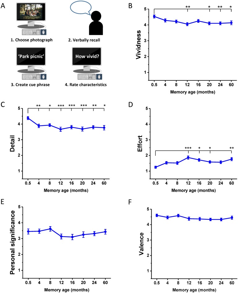
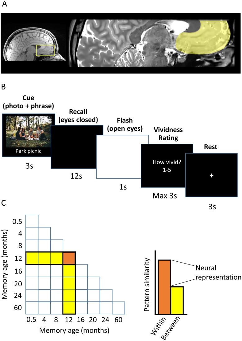
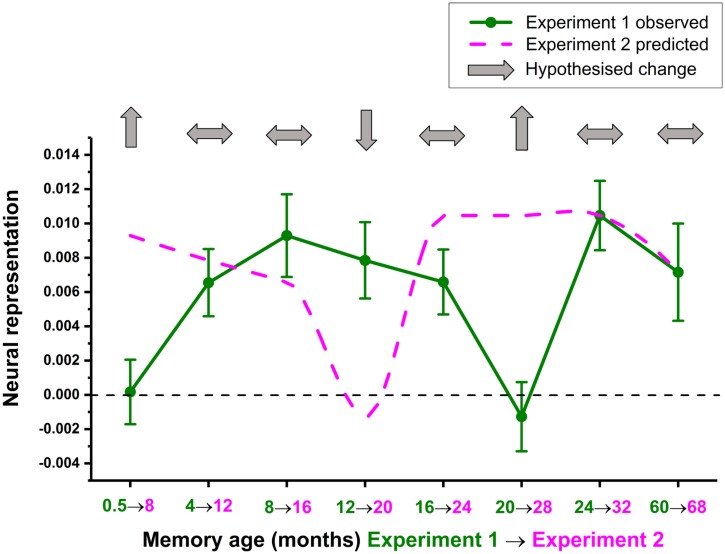
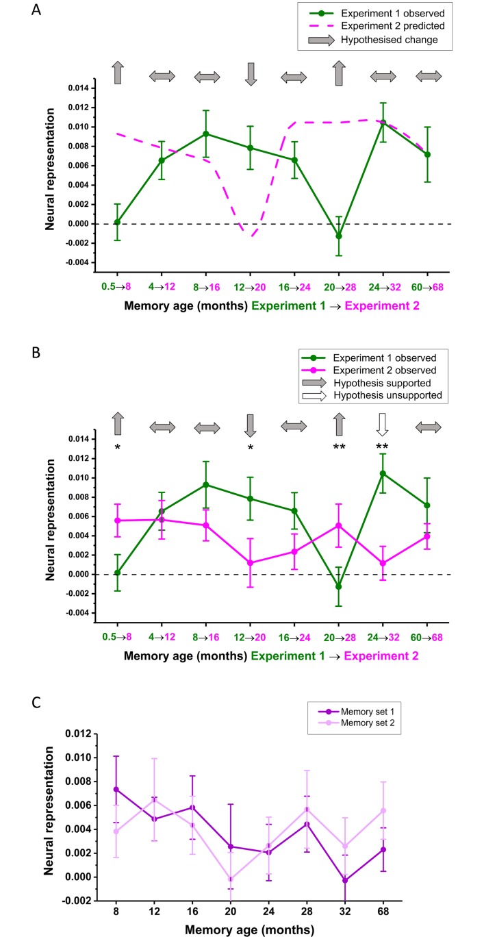
Systems-level consolidation refers to the time-dependent reorganisation of memory traces in the neocortex, a process in which the ventromedial prefrontal cortex (vmPFC) has been implicated. Capturing the precise temporal evolution of this crucial process in humans has long proved elusive. Here, we used multivariate methods and a longitudinal functional magnetic resonance imaging (fMRI) design to detect, with high granularity, the extent to which autobiographical memories of different ages were represented in vmPFC and how this changed over time. We observed an unexpected time course of vmPFC recruitment during retrieval, rising and falling around an initial peak of 8-12 months, before reengaging for older 2- and 5-year-old memories. This pattern was replicated in 2 independent sets of memories. Moreover, it was further replicated in a follow-up study 8 months later with the same participants and memories, for which the individual memory representations had undergone their hypothesised strengthening or weakening over time. We conclude that the temporal engagement of vmPFC in memory retrieval seems to be nonmonotonic, revealing a complex relationship between systems-level consolidation and prefrontal cortex recruitment that is unaccounted for by current theories.


Conflict of interest statement

The authors have declared that no competing interests exist.

Figures

Fig 1
Fig 1. Memory harvesting and subjective ratings.
(A) Schematic of the interview in which the autobiographical memories were harvested. Participants recalled a memory—which was cued by a personal photograph—chose a phrase to help remind them of this memory during the subsequent scanner task, and rated its characteristics. (B-F) Subjective ratings (means +/− 1 SEM; see also means and SDs in Table A in S1 Table, and S1 Data for individual ratings across both sets of memories) of memory characteristics at each time period for Experiment 1, averaged across the two sets of memories. Ratings were on a scale of 1 to 5, in which 1 was low and 5 was high. For emotional valence: 1–2 = negative, 3 = neutral, 4–5 = positive. * p < 0.05, ** p < 0.01, *** p < 0.001.
Fig 2
Fig 2. Objective scores for memory details.
The mean +/− 1 SEM (see also means and SDs in Table B in S1 Table, and S2 Data for individual participant scores) number of internal and external details at each time period, averaged across the two sets of autobiographical memories.
Fig 3
Fig 3. Experimental details.
(A) The vmPFC is highlighted on an example participant’s structural MRI scan. (B) The timeline of an example trial from the scanning task. (C) Graphical illustration of the neural representation score calculation using RSA. The neural pattern similarity across trials recalling the same memory (orange) minus the mean pattern similarity between that memory and other memories (yellow) generates a ‘neural representation’ score. A score significantly higher than 0 indicates a neural pattern distinct to that memory is present in the vmPFC. RSA, representational similarity analysis; vmPFC, ventromedial prefrontal cortex.
Fig 4
Fig 4. fMRI results of Experiment 1.
(A) Mean +/− 1 SEM neural representation scores at each time point averaged across the two sets of memories. Asterisks above the dotted line indicate detectability of memories in vmPFC at each time point. Asterisks above the solid line indicate significant increases in memory representations compared to the most recent (0.5 M old) memories. * p < 0.05, ** p < 0.01, *** p < 0.001. See S1 Fig for the underlying RSM and S2 Fig for a box plot distribution of these data. (B) Neural representation scores at each time point plotted separately for the two sets of autobiographical memories. (C) Neural representation scores when using a single identically aged memory as a baseline. See S3 Data for individual participant scores. fMRI, functional magnetic resonance imaging; RSM, representational similarity matrix; vmPFC, ventromedial prefrontal cortex.
Fig 5
Fig 5. Predicted fMRI changes 8 months later in Experiment 2.
Predicted changes in the neural representations of individual autobiographical memories after 8 months (pink dotted line), based on shifting the original observed data forwards by 2 time points for the 16 participants from Experiment 1 (green line) who returned for Experiment 2 (see S4 Data for original and predicted values). Light grey arrows indicate the hypotheses. fMRI, functional magnetic resonance imaging.
Fig 6
Fig 6. Memory recall and subjective ratings.
(A) Schematic of the interview in which participants recalled an autobiographical memory using their previously chosen photograph and cue phrase and rated its characteristics. (B-F) Subjective ratings (means +/− 1 SEM; see also means and SDs in Table A in S1 and S3 Tables) of memory characteristics at each time period for Experiment 1 (blue line, n = 16 participants) and how the ratings of the same memories differed 8 months later during Experiment 2 (red line, the same n = 16 participants) averaged across the two sets of memories in both cases (see S5 Data for individual ratings across both sets of memories). Ratings were on a scale of 1 to 5, in which 1 was low and 5 was high. For emotional valence: 1–2 = negative, 3 = neutral, 4–5 = positive. Asterisks indicate significant differences in memory ratings between Experiments 1 and 2; * p < 0.05, ** p < 0.01, *** p < 0.001.
Fig 7
Fig 7. Objective scores for memory details over time.
The mean +/− 1 SEM (see also means and SDs in Table B in S1 and S3 Tables) number of internal and external details at each time period for Experiment 1 (blue bars, n = 16 participants) and Experiment 2 (red bars, the same n = 16 participants), averaged across the two sets of autobiographical memories (see S6 Data for individual participant scores).
Fig 8
Fig 8. fMRI results of Experiment 2.
(A) A reminder of the hypothesised changes in neural representations from Experiment 1 (green line) to Experiment 2 (pink line, reprinted from Fig 5). (B) Mean +/− 1 SEM neural representation scores at each time point averaged across the two sets of memories for Experiment 2 (pink line, n = 16 participants) compared to the same memories from 8 months previously (green line, the same n = 16 participants). Light grey and white arrows indicate supported and unsupported hypotheses, respectively; * p < 0.05, ** p < 0.01. (C) Neural representation scores at each time point for Experiment 2, plotted separately for the two sets of autobiographical memories. See S7 Data for individual participant scores. fMRI, functional magnetic resonance imaging.

References

    1. Kandel ER. The molecular biology of memory storage: a dialogue between genes and synapses. Science. 2001;294(5544):1030–8. 10.1126/science.1067020 - DOI - PubMed
    1. Pastalkova E, Serrano P, Pinkhasova D, Wallace E, Fenton AA, Sacktor TC. Storage of spatial information by the maintenance mechanism of LTP. Science. 2006;313(5790):1141–4. 10.1126/science.1128657 - DOI - PubMed
    1. Runyan JD, Dash PK. Inhibition of hippocampal protein synthesis following recall disrupts expression of episodic-like memory in trace conditioning. Hippocampus. 2005;15(3):333–9. 10.1002/hipo.20055 - DOI - PubMed
    1. Guzowski JF. Insights into immediate-early gene function in hippocampal memory consolidation using antisense oligonucleotide and fluorescent imaging approaches. Hippocampus. 2002;12(1):86–104. 10.1002/hipo.10010 - DOI - PubMed
    1. Morris RG, Moser EI, Riedel G, Martin SJ, Sandin J, Day M, et al. Elements of a neurobiological theory of the hippocampus: the role of activity-dependent synaptic plasticity in memory. Phil Trans R Soc Lond B, Biol Sci. 2003;358(1432):773–86. - PMC - PubMed

Publication types

LinkOut - more resources