Review
. 2018 Jul 1;11(7):1125.
doi: 10.3390/ma11071125.

Therapeutic Contact Lenses with Polymeric Vehicles for Ocular Drug Delivery: A Review

Affiliations
Review

Therapeutic Contact Lenses with Polymeric Vehicles for Ocular Drug Delivery: A Review

Seung Woo Choi et al. Materials (Basel). .

Abstract

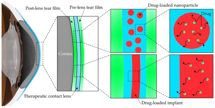
The eye has many barriers with specific anatomies that make it difficult to deliver drugs to targeted ocular tissues, and topical administration using eye drops or ointments usually needs multiple instillations to maintain the drugs’ therapeutic concentration because of their low bioavailability. A drug-eluting contact lens is one of the more promising platforms for controllable ocular drug delivery, and, among various manufacturing methods for drug-eluting contact lenses, incorporation of novel polymeric vehicles with versatile features makes it possible to deliver the drugs in a sustained and extended manner. Using the diverse physicochemical properties of polymers for nanoparticles or implants that are selected according to the characteristics of drugs, enhancement of encapsulation efficiency and prolonged drug release are possible. Even though therapeutic contact lenses with polymeric vehicles allow us to achieve sustained ocular drug delivery, drug leaching during storage and distribution and the possibility of problems related to surface roughness due to the incorporated vehicles still need to be discussed before application in a real clinic. This review highlights the overall trends in methodology to develop therapeutic contact lenses with polymeric vehicles and discusses the limitations including comparison to cosmetically tinted soft contact lenses.

Keywords: contact lenses; drug-eluting; nanoparticles; ocular drug delivery; polymeric vehicles.


Conflict of interest statement

The authors declare no conflict of interest.

Figures

Figure 1
Figure 1
Schematic of drug release onto the ocular surface from drug-eluting therapeutic contact lens. Typical polymeric vehicles for therapeutic contact lenses include drug-loaded polymeric nanoparticles and drug-loaded polymeric implants inside contact lenses.
Figure 2
Figure 2
(a) Schematic of a therapeutic contact lens embedded with timolol–PGT nanoparticles; (b) Transmittance spectra of silicone control and PGT nanoparticle-embedded silicone hydrogel. The inset shows a photograph of the PGT nanoparticle-embedded silicone hydrogel; (c) Cumulative drug release profile from timolol–PGT nanoparticle-loaded hydrogels with 100 and 200 μm thickness, respectively; (d) Pharmacodynamic profile of timolol–PGT nanoparticle-loaded contact lenses in beagle dogs, which was expressed as the difference between the intraocular pressure (IOP) of the untreated (OS) and the treated eye (OD). The IOP-lowering effect due to timolol–PGT nanoparticle-loaded contact lenses was seen on day 2 and day 3. Reprinted with permission from [28].
Figure 3
Figure 3
(a) Schematic of a therapeutic contact lens embedded with nanoparticles composed of an outer shell (polyethylene glycol), an inner shell (poly-hydroxyethylmethacrylate) and a core (polycaprolactone); (b) Photographs of transparent nanoparticle-loaded hydrogels with different nanoparticle loadings; (c) Frequency-dependent storage moduli of control hydrogel and nanoparticle-embedded hydrogel; (d) Cumulative drug release profiles from free loteprednol etabonate (LPE)-dispersed hydrogel (●), LPE-loaded nanoparticle (▲), and LPE-loaded nanoparticle-embedded hydrogel(■). Reprinted with permission from [29].
Figure 4
Figure 4
(a) Schematic of a therapeutic contact lens embedded with sandwiched polymeric implant (left, middle) and photograph (right) of a ciprofloxacin-loaded PLGA implant-embedded poly-HEMA-contact lens with a 5-mm clear central zone; (b) Cumulative drug release profiles for 100 days from free fluorescein powder, fluorescein-PLGA films, fluorescein-coated poly-HEMA-contact lens and poly-HEMA-contact lens embedded with fluorescein-PLGA films; (c) Ciprofloxacin release from PLGA implant-embedded contact lenses in vitro. Reprinted with permission from [63]; (d) SEM images (X190) of econazole-PLGA films within prototype contact lenses made of poly-HEMA hydrogel in before drug release (up) and in after 10 days of release (down). Reprinted with permission from [64]; (e) Concentration of latanoprost in aqueous humor after topical instillation of latanoprost solution (50 μg/mL); (f) Concentrations of latanoprost in aqueous humor in rabbits wearing latanoprost-eluting contact lenses. Reprinted with permission from [65].
Figure 5
Figure 5
(a) Schematic of therapeutic contact lenses embedded with frontally-located implant; (b) Schematic of the fabricating process of contact lenses embedded with an HA-laden implant (Blank arrow: inner margin of the implant, white arrow: outer margin of the implant); (c) hyaluronic acid (HA) release profile in tear fluid after wearing sterilized HA implant contact lenses and soaked contact lenses on rabbit eyes; (d) changes of ocular staining levels in dry eye disease-induced rabbit eyes by benzalkonium chloride treated with HA-implant contact lenses. Reprinted with permission from [66].
Figure 6
Figure 6
(a) Schematic of therapeutic contact lenses embedded with a sandwiched polymeric implant with timolol-encapsulated ethyl cellulose nanoparticles; schematic showing the process of fabricating contact lenses by (b) fabrication of partially UV-polymerized contact lenses and then (c) implantation of a ring implant between partially polymerized contact lenses; (d) Photograph of implant-embedded contact lens on a male mold showing a clear 6 mm central aperture and a translucent ring (White arrow: inner margin of the implant, black arrow: outer margin of the implant); (e) Change in intraocular pressure (IOP) in rabbits treated with eye drop and timolol-laden implant-embedded contact lens. Reprinted with permission from [62].
Figure 7
Figure 7
SEM images of (a) the surface of pure poly-HEMA contact lens; (b) the surface of nanoparticle-laden poly-HEMA contact lens with visible nanoparticles (red circle). Reprinted with permission from [30]; (c) Timolol-eluting profiles from drug-soaked (black line), molecularly imprinted (red line) and nanodiamond nanogel-embedded contact lenses (blue line), showing lysozyme-triggered drug release. Reprinted with permission from [125].

References

    1. Gaudana R., Ananthula H.K., Parenky A., Mitra A.K. Ocular drug delivery. AAPS J. 2010;12:348–360. doi: 10.1208/s12248-010-9183-3. - DOI - PMC - PubMed
    1. Bachu R.D., Chowdhury P., Al-Saedi Z.H.F., Karla P.K., Boddu S.H.S. Ocular drug delivery barriers-role of nanocarriers in the treatment of anterior segment ocular diseases. Pharmaceutics. 2018;10:28. doi: 10.3390/pharmaceutics10010028. - DOI - PMC - PubMed
    1. Lang J.C. Ocular drug delivery conventional ocular formulations. Adv. Drug Deliv. Rev. 1995;16:39–43. doi: 10.1016/0169-409X(95)00012-V. - DOI
    1. Janagam D.R., Wu L., Lowe T.L. Nanoparticles for drug delivery to the anterior segment of the eye. Adv. Drug Deliv. Rev. 2017;122:31–64. doi: 10.1016/j.addr.2017.04.001. - DOI - PMC - PubMed
    1. Novack G.D. Ophthalmic drug delivery: Development and regulatory considerations. Clin. Pharmacol. Ther. 2009;85:539–543. doi: 10.1038/clpt.2008.297. - DOI - PubMed