. 2018 Jul 1;57(4):350-356.
doi: 10.30802/AALAS-JAALAS-17-000163. Epub 2018 Jul 2.

Behavioral Assessment of Vision in Pigs

Affiliations

Behavioral Assessment of Vision in Pigs

Francesca Barone et al. J Am Assoc Lab Anim Sci. .

Abstract

Swine (Sus scrofa) are often the 'gold standard' laboratory animal for ophthalmology research due to the anatomic and physiologic similarities between the porcine and human eye and retina. Despite the importance of this model, few tools for behavioral vision assessment in pigs are available. The aim of this study was to identify and validate a feasible and reproducible behavioral test to assess vision in a pig model of photoreceptor degeneration. In addition, a robust behavioral test will reduce stress and enhance enrichment by allowing animals opportunities for environmental exploration and by reducing the number of invasive experimental procedures. Two distinct behavioral approaches were tested: the obstacle-course test and temperament test. In the obstacle-course test, pigs were challenged (after an initial training period) to navigate a 10-object obstacle course; time and the number of collisions with the objects were recorded. In the temperament test, the time needed for pigs to complete 3 different tasks (human-approach, novel-object, and open-door tests) was recorded. The obstacle-course test revealed significant differences in time and number of collisions between swine with vision impairment and control animals, and the training period proved to be pivotal to avoid bias due to individual animal characteristics. In contrast, the temperament test was not altered by vision impairment but was validated to measure stress and behavioral alterations in laboratory pigs undergoing experimental procedures, thus achieving marked refinement of the study.


Figures
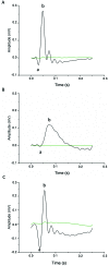
Figure 1.
Figure 1.
Representative ff-ERG waves. (A) Light-adapted 3.0 ERG, b waves correspond to bipolar cell activation after cones respond to light stimuli. (B) Dark-adapted 0.01 ERG, b waves correspond to bipolar cell activation after rods respond to light stimuli. (C) Dark-adapted 3.0 ERG, b waves correspond to bipolar cell activation after all photoreceptors response to light stimuli. Because dark-adapted ERG are recorded sequentially without further dark adaptation, only the 0.01 ERG is a fully dark-adapted response. Black waves correspond to a representative animal from group C; green waves to a representative of the IAA-treated group.
Figure 2.
Figure 2.
Obstacle course test. Graphic representation of the 4 obstacle configurations from above and side and front views of the obstacles.
Figure 3.
Figure 3.
Time line for (A) obstacle course test and (B) temperament testing. m, Monday; w, Wednesday; f, Friday; HAT, humane approach test; NOT, novel object test; ODT, open door test.
Figure 4.
Figure 4.
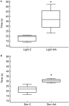
Obstacle course test. The box plots indicate the median and interquartile range. (A) Time to complete the course under the light condition. (B) Time to complete the course under dim conditions. C, control group (n = 5); IAA, Iodoacetic Acid group n = 9. *, P < 0.05 (Mann–Whitney U test) between C and IAA groups; no differences were detected between test conditions.
Figure 5.
Figure 5.
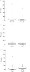
Temperament Test. Box plot median interquartile range (n = 8). (A) Human approach test. (B) Novel object test. (C) Open door test. □, Out-of-range values; ○, outliers.

References

    1. Antognazza MR, Di Paolo M, Ghezzi D, Mete M, Di Marco S, Maya-Vetencourt JF, Maccarone R, Desii A, Di Fonzo F, Bramini M, Russo A, Laudato L, Donelli I, Cilli M, Freddi G, Pertile G, Lanzani G, Bisti S, Benfenati F. 2016. Characterization of a polymer-based, fully organic prosthesis for implantation into the subretinal space of the rat. Adv Healthc Mater 5:2271–2282. 10.1002/adhm.201600318. - DOI - PubMed
    1. Augsburger AS, Haag V, Leuillet S, Legrand JJ, Forster R. 2012. Recording of the full-field electroretinogram in minipigs. Vet Ophthalmol 15 Suppl 2:84–93. 10.1111/j.1463-5224.2012.01052.x. - DOI - PubMed
    1. Brunjes PC, Feldman S, Osterberg SK. 2016. The pig olfactory brain: a primer. Chem Senses 41:415–425. 10.1093/chemse/bjw016. - DOI - PMC - PubMed
    1. Chandler MJ, Smith PJ, Samuelson DA, MacKay EO. 1999. Photoreceptor density of the domestic pig retina. Vet Ophthalmol 2:179–184. 10.1046/j.1463-5224.1999.00077.x. - DOI - PubMed
    1. Curcio CA, Sloan KR, Kalina RE, Hendrickson AE. 1990. Human photoreceptor topography. J Comp Neurol 292:497–523. 10.1002/cne.902920402. - DOI - PubMed

Publication types