

Hearing and orally mimicking different acoustic-semantic categories of natural sound engage distinct left hemisphere cortical regions
- PMID: 29966815
- PMCID: PMC6461214
- DOI: 10.1016/j.bandl.2018.05.002
Hearing and orally mimicking different acoustic-semantic categories of natural sound engage distinct left hemisphere cortical regions
Abstract
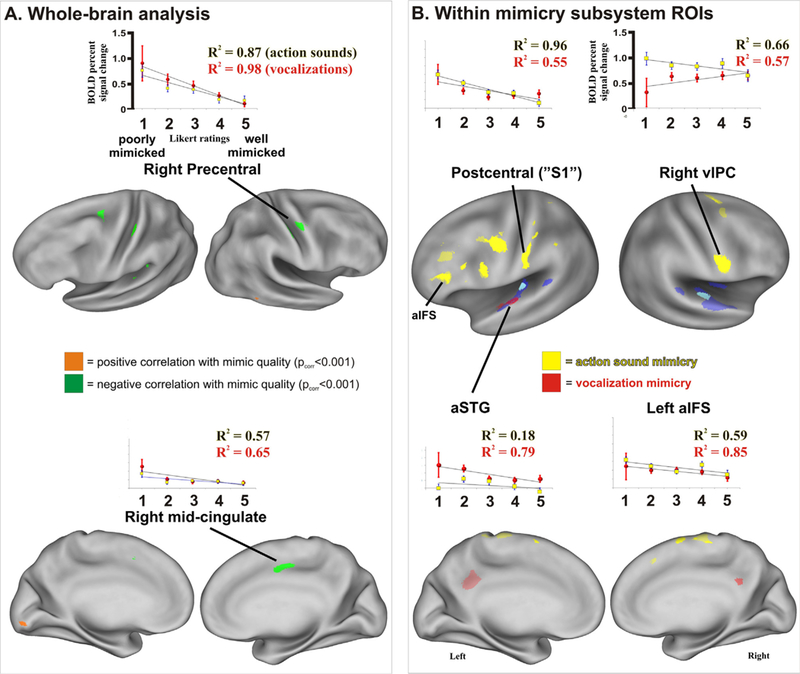
Oral mimicry is thought to represent an essential process for the neurodevelopment of spoken language systems in infants, the evolution of language in hominins, and a process that could possibly aid recovery in stroke patients. Using functional magnetic resonance imaging (fMRI), we previously reported a divergence of auditory cortical pathways mediating perception of specific categories of natural sounds. However, it remained unclear if or how this fundamental sensory organization by the brain might relate to motor output, such as sound mimicry. Here, using fMRI, we revealed a dissociation of activated brain regions preferential for hearing with the intent to imitate and the oral mimicry of animal action sounds versus animal vocalizations as distinct acoustic-semantic categories. This functional dissociation may reflect components of a rudimentary cortical architecture that links systems for processing acoustic-semantic universals of natural sound with motor-related systems mediating oral mimicry at a category level. The observation of different brain regions involved in different aspects of oral mimicry may inform targeted therapies for rehabilitation of functional abilities after stroke.
Keywords: Acoustic communication; Acoustic-semantic categories; Categorical perception; Echo-mirror neuron system; Language evolution; Sound symbolism; Stroke rehabilitation; fMRI.
Published by Elsevier Inc.
Figures
Similar articles
-
Different categories of living and non-living sound-sources activate distinct cortical networks.Neuroimage. 2009 Oct 1;47(4):1778-91. doi: 10.1016/j.neuroimage.2009.05.041. Epub 2009 May 22. Neuroimage. 2009. PMID: 19465134 Free PMC article.
-
Cortical representation of natural complex sounds: effects of acoustic features and auditory object category.J Neurosci. 2010 Jun 2;30(22):7604-12. doi: 10.1523/JNEUROSCI.0296-10.2010. J Neurosci. 2010. PMID: 20519535 Free PMC article.
-
Cortical networks representing object categories and high-level attributes of familiar real-world action sounds.J Cogn Neurosci. 2011 Aug;23(8):2079-101. doi: 10.1162/jocn.2010.21570. Epub 2010 Sep 2. J Cogn Neurosci. 2011. PMID: 20812786
-
Auditory object perception: A neurobiological model and prospective review.Neuropsychologia. 2017 Oct;105:223-242. doi: 10.1016/j.neuropsychologia.2017.04.034. Epub 2017 Apr 30. Neuropsychologia. 2017. PMID: 28467888 Free PMC article. Review.
-
Neural processing of natural sounds.Nat Rev Neurosci. 2014 Jun;15(6):355-66. doi: 10.1038/nrn3731. Nat Rev Neurosci. 2014. PMID: 24840800 Review.
Cited by
-
Chinese-English bilinguals show linguistic-perceptual links in the brain associating short spoken phrases with corresponding real-world natural action sounds by semantic category.Lang Cogn Neurosci. 2021;36(6):773-790. doi: 10.1080/23273798.2021.1883073. Epub 2021 Feb 17. Lang Cogn Neurosci. 2021. PMID: 34568509 Free PMC article.
-
Electrophysiological Evidence of Early Cortical Sensitivity to Human Conspecific Mimic Voice as a Distinct Category of Natural Sound.J Speech Lang Hear Res. 2020 Oct 16;63(10):3539-3559. doi: 10.1044/2020_JSLHR-20-00063. Epub 2020 Sep 16. J Speech Lang Hear Res. 2020. PMID: 32936717 Free PMC article.
-
Meta-Analyses Support a Taxonomic Model for Representations of Different Categories of Audio-Visual Interaction Events in the Human Brain.Cereb Cortex Commun. 2021 Jan 18;2(1):tgab002. doi: 10.1093/texcom/tgab002. eCollection 2021. Cereb Cortex Commun. 2021. PMID: 33718874 Free PMC article.
References
-
- Arbib MA (2005). From monkey-like action recognition to human language: an evolutionary framework for neurolinguistics. Behavioral and Brain Sciences, 28, 105–124 (discussion 125–167). - PubMed
-
- Asano M, Imai M, Kita S, Kitajo K, Okada H, &Thierry G (2015). Sound symbolism scaffolds language development in preverbal infants. Cortex, 63, 196–205. - PubMed
-
- Baddeley AD (1986). Working memory. In Gazzaniga MS (Ed.). Principles of neuroscience. MIT Press: Clarendon Press.
-
- Belin P, Zatorre RJ, Lafaille P, Ahad P, & Pike B (2000). Voice-selective areas in human auditory cortex. Nature, 403, 309–312. - PubMed
Publication types
MeSH terms
Grants and funding
LinkOut - more resources
Full Text Sources
Other Literature Sources
