. 2018 Oct:36:12-20.
doi: 10.1016/j.anndiagpath.2018.06.002. Epub 2018 Jun 18.

MiR-155 deletion reduces ischemia-induced paralysis in an aortic aneurysm repair mouse model: Utility of immunohistochemistry and histopathology in understanding etiology of spinal cord paralysis

Affiliations

MiR-155 deletion reduces ischemia-induced paralysis in an aortic aneurysm repair mouse model: Utility of immunohistochemistry and histopathology in understanding etiology of spinal cord paralysis

Hamdy Awad et al. Ann Diagn Pathol. 2018 Oct.

Abstract

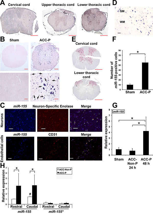
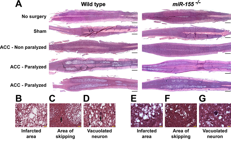
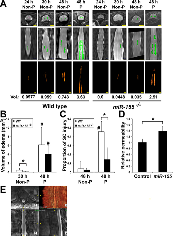
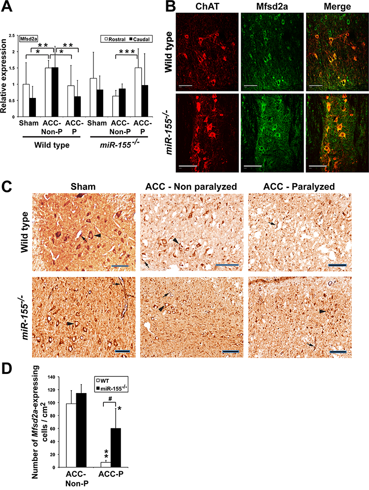
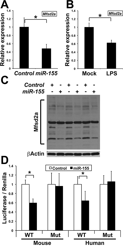
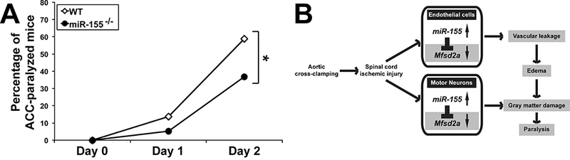
Spinal cord paralysis is relatively common after surgical repair of thoraco-abdominal aortic aneurysm (TAAA) and its etiology is unknown. The present study was designed to examine the histopathology of the disease and investigate whether miR-155 ablation would reduce spinal cord ischemic damage and delayed hindlimb paralysis induced by aortic cross-clamping (ACC) in our mouse model. The loss of locomotor function in ACC-paralyzed mice correlated with the presence of extensive gray matter damage and central cord edema, with minimal white matter histopathology. qRTPCR and Western blotting showed that the spinal cords of wild-type ACC mice that escaped paralysis showed lower miR-155 expression and higher levels of transcripts encoding Mfsd2a, which is implicated in the maintenance of blood-brain barrier integrity. In situ based testing demonstrated that increased miR-155 detection in neurons was highly correlated with the gray matter damage and the loss of one of its targets, Mfsd2a, could serve as a good biomarker of the endothelial cell damage. In vitro, we demonstrated that miR-155 targeted Mfsd2a in endothelial cells and motoneurons and increased endothelial cell permeability. Finally, miR-155 ablation slowed the progression of central cord edema, and reduced the incidence of paralysis by 40%. In sum, the surgical pathology findings clearly indicated that the epicenter of the ischemic-induced paralysis was the gray matter and that endothelial cell damage correlated to Mfsd2a loss is a good biomarker of the disease. MiR-155 targeting therefore offers new therapeutic opportunity for edema caused by traumatic spinal cord injury and diagnostic pathologists, by using immunohistochemistry, can clarify if this mechanism also is important in other ischemic diseases of the CNS, including stroke.

Keywords: Histopathology; Immunohistochemistry; Ischemia; Spinal cord; microRNA.


Conflict of interest statement

Potential conflicts of interest

Nothing to report.

Figures

Fig. 1.
Fig. 1.
Up-regulation of miR-155 expression in SC of ACC-P WT mice. (A) In situ hybridization showing miR-155 expression on three cross sections of spinal cord of the same mouse 48 h following ACC. Scale bars = 400 μm. (B) Representative in situ hybridization for miR-155 on cross sections of the lower thoracic part of SC (44–48 h following ACC) of four sham and four ACC-P mice. Right lower panel: black arrows point to miR-155-positive endothelial cells of blood vessels. Counterstain is nuclear fast red. Top panels: scale bars = 200 μm. Lower panels: scale bars = 40 μm. (C) MiR-155 expression in neurons of WT mice was assessed using an antibody to Neuron-Specific Enolase neuronal marker. MiR-155 expression in endothelial cells was assessed using an antibody to CD31. Yellow: co-expression. Scale bars = 125 μm. (D) MiR-155 expression in wild type ACC-Paralyzed mice is primarily detected in spinal cord gray matter. WM, White matter. GM, Gray matter. Scale bar = 100 μm. (E) NeuN expression in ventral horns of lower thoracic spinal cord following ACC; note loss of expression corresponds to high miR-155 expression. Scale bars = 400 μm. (F) Number (Mean + SD) of miR-155 positive cells on cross-sections of the thoracic and lumbar regions of SC of sham (n = 4) and ACC-P (n = 4) mice 44–48 h after ACC (3 counts/ mouse). *, P < 0.0042. (G) MiR-155 expression (Mean + SD) in the caudal region of the spinal cord of sham (n = 5), 24 h ACC-Non-P (n = 5) and 44–48 h ACC-P (n = 4) WT mice was determined by qRTPCR. *, P < 0.0459. Values were normalized to sham. (H) MiR-155 and miR-155* expression (Mean + SD) in the rostral and caudal regions of SC of ACC-Non-P (n = 4) and ACC-P (n = 7) WT mice was determined by qRT-PCR 44–48 h after ACC. *,P < 0.031;#, caudal ACC-Non-P different from rostral ACC-Non-P, P < 0.0016. Values were normalized to rostral ACC-Non-P miR-155. (For interpretation of the references to colour in this figure legend, the reader is referred to the web version of this article.)
Fig. 2.
Fig. 2.
Correlation of spinal cord histological damage with paralysis in WT and miR-155−/− ACC mice. (A) Representative images of H&E-stained, longitudinal sections through the ventral horns of spinal cord of no-surgery, sham, ACC-Non-P and ACC-P WT and miR-155−/− mice, 44–48 h following ACC. Rostral is on the right side. Scale bars = 1 mm. (B, E) High definition of the large area of infarct from a WT (B) and a miR-155−/− (E) ACC-P mouse. Scale bars = 20 μm. (C, F) Skipping area [black arrow in (C)] in the same spinal cords as in (B) and (E), respectively, located between two infarcted areas. Scale bars = 20 μm. (D, G) Presence of vacuolated neurons in the same spinal cords as in (B) and (E), respectively. Scale bars = 20 μm.
Fig. 3.
Fig. 3.
MiR-155 interferes with the development of central cord edema (CCE) following ACC. (A) Axial (top panels) and coronal (middle panels) images of T2 weighted WT and miR-155−/− mouse thoracic and lumbar spinal cords as seen by MRI. The volumes (Vol., in mm3) of increased T2 signal felt to represent CCE (bottom panels) was calculated by outlining areas of increased T2 signal. Scale bars = 2 mm. (B) Volume (Mean + SD) of edema in WT and miR-155−/− ACC mice. *, P < 0.0455;#, 48 h paralyzed different from corresponding 30 h Non-P, P < 0.0049. At 30 h: WT, n = 6; miR-155−/−, n = 8. At 48 h: WT, n = 8; miR-155−/−, n = 6. (C) Proportion of spinal cord injury (Mean + SD), defined as the ratio of ischemic area/total, in WT (Non-P: n = 6; P: n = 6) and miR-155−/− (Non-P: n = 5; P: n = 4) mice 44–48 h after ACC (3 counts/mouse). *, P < 0.0039;#, WT paralyzed different from WT non-paralyzed, P < 0.000046. (D) Relative permeability of a monolayer of mouse endothelial cells to FITC-Dextran 48 h after transfection with either a control pre-miRNA (Control) or premiR-155 (miR-155). *, P < 0.0000036 (n = 11 in both groups). Values were normalized to Control. (E) Spinal cord edema in a TAAA repair patient experiencing uni-lateral lower extremity paresis 3–4 days following TAAA repair surgery was analyzed using a Three Tesla MRI of the thoracic spine utilizing a Siemens Verio scanner with a TR (time to repetition pulse) of 872 ms and TE (time to echo) of 7.8 ms. Three Tesla MRI findings of faint increased T2 signal within central gray matter of the lower thoracic SC extending from T10 to T12 into the conus medullaris and cauda equina, consistent with CCE (volume = 2.96 cm3). Scale bar = 5 mm (top right panel). Scale bars = 2 mm (three other panels).
Fig. 4.
Fig. 4.
Mfsd2a expression is affected by ischemic conditions. (A) Mfsd2a relative expression (Mean + SD) 44–48 h after ACC in the rostral and caudal parts of spinal cord of WT sham (n = 9), ACC-Non-P (n = 5) and ACC-P (n = 7) mice, as well as of miR-155−/− sham (n = 5), ACC-Non-P (n = 5) and ACC-P (n = 10) mice, as shown by qRTPCR. *, P < 0.0668; **, P < 0.0361; ***, P < 0.00096. Values were normalized to WT rostral sham. (B) Mfsd2a (green) and ChAT (red), a marker of spinal cord moto-neurons, co-localize (yellow), in both sham WT and sham miR-155−/− mice, as shown by immunohistochemistry. Scale bars = 100 μm. (C) Mfsd2a-expressing cells in spinal cord of WT and miR-155−/− mice. Arrow heads: motoneurons. Arrows: endothelial cells. Scale bars = 100 μm. (D) Number of Mfsd2a-expressing endothelial cells and neurons (Mean + SD) 44–48 h after ACC in spinal cord of the same WT and miR-155−/− mice as in Fig. 3C (3 counts/ mouse). ** and *, ACC-P different from corresponding ACC-Non P; **, P < 0.000093; *, P < 0.0312;#,P < 0.0027. (For interpretation of the references to colour in this figure legend, the reader is referred to the web version of this article.)
Fig. 5.
Fig. 5.
MiR-155 targets transcripts encoding Mfsd2a. (A) Mfsd2a relative expression (Mean + SD), as determined by qRT-PCR, in mouse endothelial cells transfected with either a control pre-miRNA (Control) or pre-miR-155 (miR-155). *, P < 0.00136 (n = 6 in both groups). Values were normalized to Control. (B) Mfsd2a relative expression (Mean + SD), as determined by qRTPCR, in mouse endothelial cells either mock-treated (Mock) or cultivated in the presence of the supernatant of RAW264.7 macrophages challenged with LPS for 24 h (LPS). *, P < 0.00005 (n = 6 in both groups). Values were normalized to Mock. (C) Western blots showing the expression of multiple Mfsd2a bands in mouse endothelial cells transfected with either a control pre-miRNA (Control) or pre-miR-155 (miR-155). Results are from three independent transfection experiments. The main, constant band is most-probably non-specific. (D) Luciferase activity (Mean + SD) produced from mouse endothelial cells transfected with a Luciferase construct containing the mouse or human Mfsd2a 3′UTR with either a wild type (WT) or mutated (Mut) miR-155 target site, along with either a pre-miR-Control (Control) or pre-miR-155 (miR-155). *, P < 0.033 (n = 4 in each group). Values were normalized to Control.
Fig. 6.
Fig. 6.
(A) MiR-155 deletion reduces the rate of paralysis two days following ACC. Percentages of paralyzed WT (n = 51) and miR-155−/− (n = 38) mice following ACC. *, P = 0.05. (B) Working model showing how miR-155 is instrumental in spreading ischemic damage within the SC. Following ACC, miR-155 targets transcripts encoding Msfd2a and other protective factors, which leads to vascular leakage, development of CCE, gray matter damage and paralysis. Mfsd2a targeting in neurons is also likely to reduce DHA influx [15], with deleterious consequences on neuron survival and reduced production of anti-inflammatory DHA-derivatives such as Neuroprotectin D1. MiR-155−/− ACC-mice retain higher levels of Mfsd2a and other protective factors, thus experiencing slower CCE development, reduced gray matter damage and paralysis. MiR-155 effects in neurons, particularly on DHA supply under ischemic conditions, remain to be studied.

References

    1. Awad H, Ramadan ME, El Sayed HF, et al. Spinal cord injury after thoracic endovascular aortic aneurysm repair. Can J Anaesth 2017;64:1218–35. - PMC - PubMed
    1. Awad H, Ankeny DP, Guan Z, et al. A mouse model of ischemic spinal cord injury with delayed paralysis caused by aortic cross-clamping. Anesthesiology 2010;113:880–91. - PMC - PubMed
    1. Smith PD, Puskas F, Meng X, et al. The evolution of chemokine release supports a bimodal mechanism of spinal cord ischemia and reperfusion injury. Circulation 2012;126:S110–7. - PubMed
    1. Hamilton JA, Brunaldi K. A model for fatty acid transport into the brain. J Mol Neurosci 2007;33:12–7. - PubMed
    1. Ben-Zvi A, Lacoste B, Kur E, et al. Mfsd2a is critical for the formation and function of the blood-brain barrier. Nature 2014;509:507–11. - PMC - PubMed