Treatment of Graf Type IIa Hip Dysplasia: A Cut-off Value for Decision Making
- PMID: 29966996
- PMCID: PMC6251377
- DOI: 10.4274/balkanmedj.2017.1150
Treatment of Graf Type IIa Hip Dysplasia: A Cut-off Value for Decision Making
Abstract
Background: The rate of spontaneous normalization in type IIa hips is reported to be high, whereas dysplsia persists or worsens in 5%-10% of cases.
Aims: To evaluate the natural course of type IIa hips using Graf’s own perspective of physiological immaturity and maturational deficit.
Study design: A single center, retrospective cohort study.
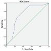
Methods: This was an institutional review board-approved retrospective review of all patients diagnosed with type IIa hip dysplasia at a single institution from 2012 to 2014. All patients included in the study had hip ultrasonography at about 6 weeks and 3 months of age. To assess reliability in α and β angles, ultrasonography measurements were carried out on the same image individually by all observers. The α and β angles were used as the main outcome measurements to evaluate hip maturation at the last follow-up. A receiver operating characteristics curve was drawn at the 3 month ultrasonography to evaluate the cut-off values for α and β angles for persistent dysplasia.
Results: Sixty-four patients and 88 affected hips (63% unilateral and 37% bilateral) were included. The mean age at diagnosis was 6.4±2.7 weeks. Fifty-four hips were type IIa(+) (physiologically immature) and 34 hips were type IIa(-) (maturational deficit) at the initial ultrasonography evaluation. Improvement to type I was seen in 52 type IIa(+) and 17 type IIa(-) hips. Receiver operating characteristic analyses showed that patients do well if the α angle was >55° (area under the curve: 0.86; p<0.001 for the left hip and area under the curve: 0.72; p=0.008 for the right hip).
Conclusion: The cut-off α angle value of 55° on initial ultrasonography should be considered to prevent future dysplasia. An α angle <55° on the initial ultrasonography was an independent predictor of worsening sonographic findings.
Keywords: Decisioion making; Graf type IIa; Hip dysplasia; Ultrasonography.
Conflict of interest statement
Figures


References
-
- Tschauner C, Fürntrath F, Saba Y, Berghold A, Radl R. Developmental dysplasia of the hip: impact of sonographic newborn hip screening on the outcome of early treated decentered hip joints-a single center retrospective comparative cohort study based on Graf's method of hip ultrasonography. J Child Orthop. 2011;5:415–24. - PMC - PubMed
-
- Puhan MA, Woolacott N, Kleijnen J, Steurer J. Observational studies on ultrasound screening for developmental dysplasia of the hip in newborns - a systematic review. Ultraschall Med. 2003;24:377–82. - PubMed
-
- Graf R. New possibilities for the diagnosis of congenital hip joint dislocation by ultrasonography. J Pediatr Orthop. 1983;3:354–9. - PubMed
MeSH terms
LinkOut - more resources
Full Text Sources
Other Literature Sources
Medical
