. 2018 Aug;30(8):1887-1905.
doi: 10.1105/tpc.18.00347. Epub 2018 Jul 2.

HEAT INDUCIBLE LIPASE1 Remodels Chloroplastic Monogalactosyldiacylglycerol by Liberating α-Linolenic Acid in Arabidopsis Leaves under Heat Stress

Affiliations

HEAT INDUCIBLE LIPASE1 Remodels Chloroplastic Monogalactosyldiacylglycerol by Liberating α-Linolenic Acid in Arabidopsis Leaves under Heat Stress

Yasuhiro Higashi et al. Plant Cell. 2018 Aug.

Abstract

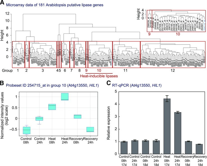
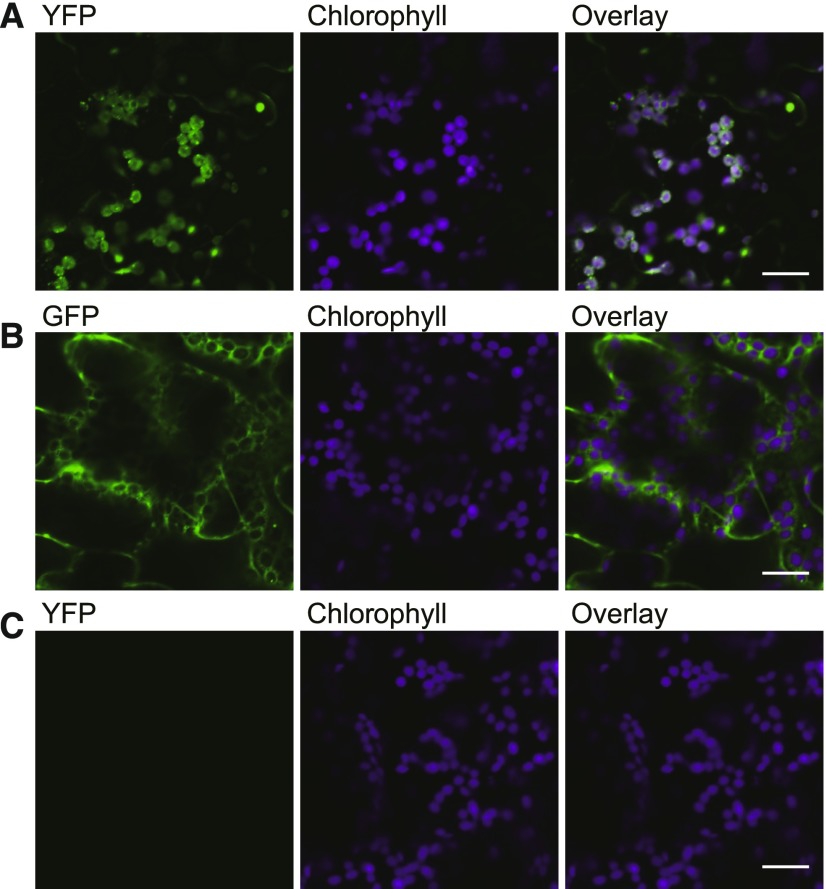
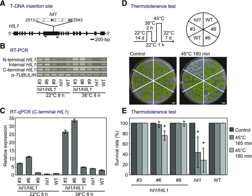
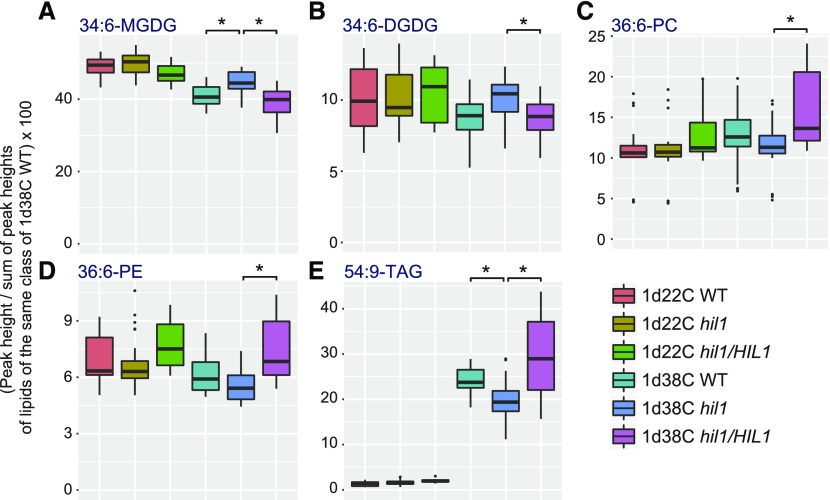
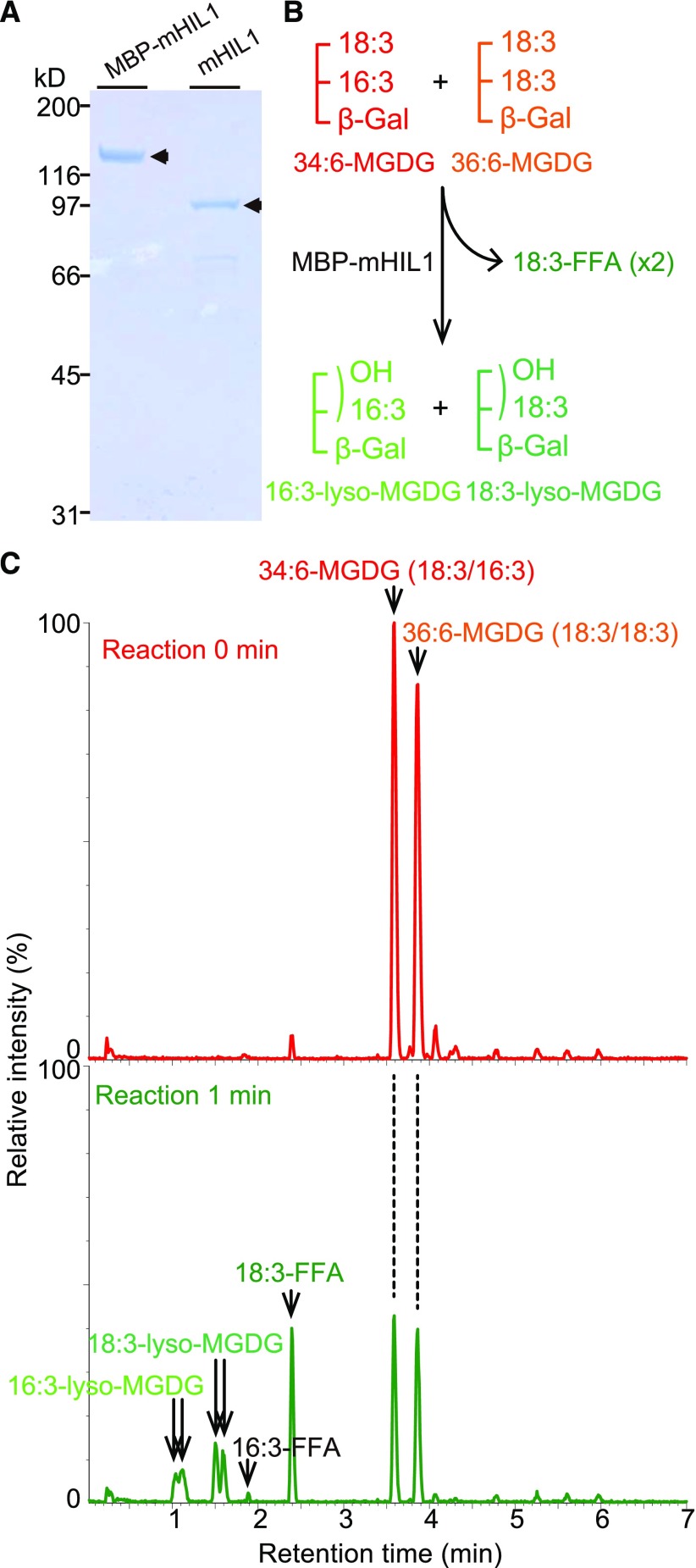
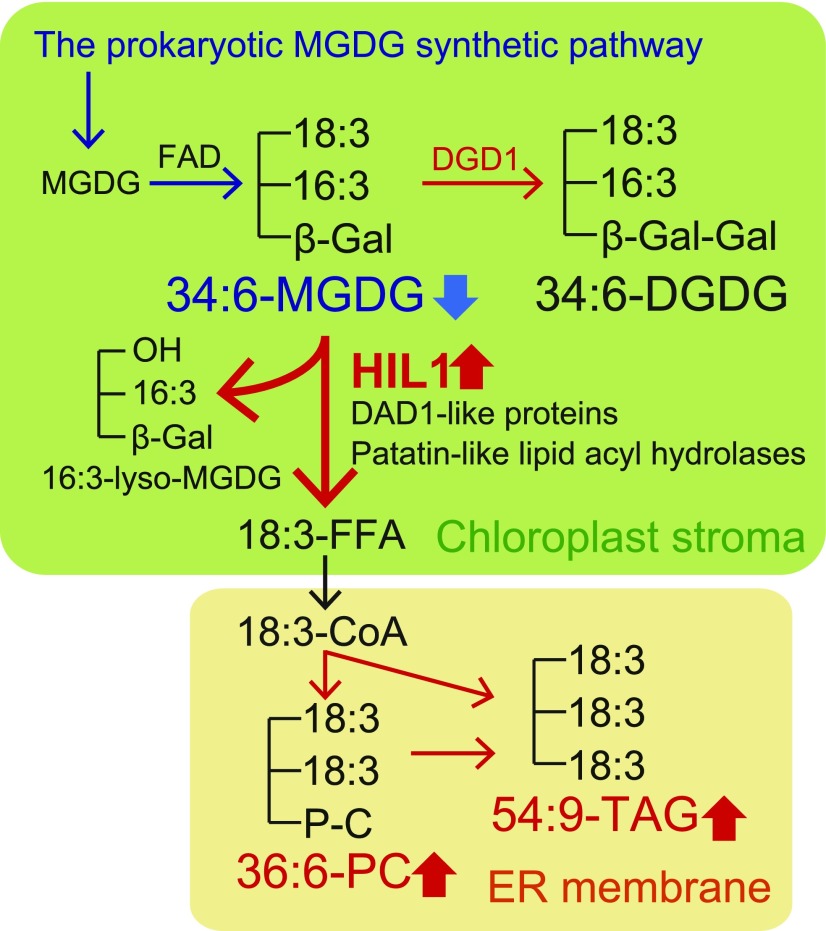
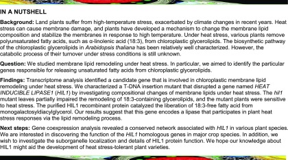
Under heat stress, polyunsaturated acyl groups, such as α-linolenate (18:3) and hexadecatrienoate (16:3), are removed from chloroplastic glycerolipids in various plant species. Here, we showed that a lipase designated HEAT INDUCIBLE LIPASE1 (HIL1) induces the catabolism of monogalactosyldiacylglycerol (MGDG) under heat stress in Arabidopsis thaliana leaves. Using thermotolerance tests, a T-DNA insertion mutant with disrupted HIL1 was shown to have a heat stress-sensitive phenotype. Lipidomic analysis indicated that the decrease of 34:6-MGDG under heat stress was partially impaired in the hil1 mutant. Concomitantly, the heat-induced increment of 54:9-triacylglycerol in the hil1 mutant was 18% lower than that in the wild-type plants. Recombinant HIL1 protein digested MGDG to produce 18:3-free fatty acid (18:3-FFA), but not 18:0- and 16:0-FFAs. A transient assay using fluorescent fusion proteins confirmed chloroplastic localization of HIL1. Transcriptome coexpression network analysis using public databases demonstrated that the HIL1 homolog expression levels in various terrestrial plants are tightly associated with chloroplastic heat stress responses. Thus, HIL1 encodes a chloroplastic MGDG lipase that releases 18:3-FFA in the first committed step of 34:6 (18:3/16:3)-containing galactolipid turnover, suggesting that HIL1 has an important role in the lipid remodeling process induced by heat stress in plants.


Figures

Figure 1.
Figure 1.
Gene Expression of Putative Lipase Genes under Heat Stress. (A) A tree diagram shows the similarity of expression patterns among the 181 Arabidopsis putative lipase genes under different heat stresses. Previous GeneChip data were used for the analysis (Higashi et al., 2015). Hierarchical clustering analysis classified the genes into 12 groups (red lines). Groups 9 and 10 (red font, enlargement in inset) contain genes (probe set IDs) induced by heat stress and returned to normal levels during temperature recovery (see Table 1 for detail). (B) Microarray data of a heat-inducible lipase (At4g13550, HIL1), which belongs to group 10 (blue font, “254715_at”) in (A). Each data point was represented by a box and whisker plot (biological replicates of n = 3 to 9). (C) The transcript levels of HIL1 were quantified by RT-qPCR. The y axis represents the relative expression level of HIL1 against “Control 08 h 17 d” calculated by the comparative CT (ΔΔCT) method. RNA content was normalized by EIF4A1 as an internal standard. Each data point expresses the mean of three experiments ± sd. The whole rosettes harvested from two to three different plants were pooled as one biological repeat.
Figure 2.
Figure 2.
HIL1 Has Relatively Unique Characteristics among the Lipase Genes. (A) Schematic representation of HIL1 structure. Regions of the putative cTP sequence, C2 domain, and lipase-like domain are shown in green, blue, and brown boxes, respectively. (B) Sequence similarity between HIL1 and DAD1-like proteins. Arabidopsis genes that exhibit homology to the lipase-like domain of HIL1 were selected. Only the lipase-like domain sequences were used to generate the amino acid sequence alignment (Supplemental Data Set 1). Twelve DAD1-like proteins formed a group (blue). HIL1 is separated from this group (purple). (C) Phylogenetic relationships among HIL1 and homologs in terrestrial plants. The phylogenetic tree was constructed from an alignment of deduced full-length amino acid sequences (Supplemental Data Set 1). The bar indicates amino acid substitutions per site. Bootstrap values (2000 replicates) > 70% are shown next to the branches.
Figure 3.
Figure 3.
Confocal Micrographs Showing Chloroplast Targeting of HIL1. The HIL1-YFP fusion protein was transiently expressed in N. benthamiana leaves. HIL1-YFP (full-length HIL1 fused with YFP) (A), free GFP (B), and p19 negative control (C). Bars = 20 μm.
Figure 4.
Figure 4.
Genomic Organization of the hil1 T-DNA Insertion Line, and the Impact of Insertional Mutation and Its Genetic Complementation on HIL1 Expression Levels and Thermotolerance. (A) Schematic representation of HIL1 (At4g13550) with a T-DNA insertion mutant (hil1) used in this work. Exons are shown as boxes. White and black triangles show left and right borders, respectively. Numbers indicate the position of the T-DNA insertion from the start codon of HIL1. The arrows indicate primers used for RT-PCR (internal HIL1) in (B). (B) RT-PCR analysis of HIL1 transcripts in the wild-type plants (WT), hil1 mutant, and hil1 complemented with the genomic sequence of HIL1 (hil1/HIL1 #3, #6, and #9 T3 lines). Total RNA was extracted from the whole rosettes of 17-d-old plants under heat stress conditions at 38°C for 8 h (38°C 8 h). As a control, plants not subjected to the stress but grown under normal conditions at 22°C were examined at the same time point (22°C 8 h). HIL1 expression levels were examined at the N-terminal, internal (interrupted by the T-DNA insertion), and C-terminal regions. α-TUBULIN was used as the control. (C) RT-qPCR of HIL1 transcripts in the wild type, hil1, hil1/HIL1 #3, #6, and #9. The y axis represents relative expression level of HIL1 against 22°C 8 h WT calculated by the comparative CT (ΔΔCT) method. RNA content was normalized by EIF4A1 as an internal standard. Each data point expresses the mean of three experiments ± sd. The whole rosettes harvested from two to three different plants were pooled as one biological repeat. Primers were designed at the C-terminal region of HIL1, which is located downstream of the T-DNA insertion site. (D) and (E) The effects of heat shock on growth of the wild-type, hil1 mutant, and hil1/HIL1-complemented plants (#3, #6, and #9 T4 lines). Fourteen-day-old plants were treated at 38°C for 2 h, 22°C for 1 h, and 45°C for 165 or 180 min (45°C 165 min and 45°C 180 min), followed by recovery at 22°C for 7 d. Plants grown continuously at 22°C were examined as the normal growth condition (Control). Photographs of the whole rosettes were taken after the recovery periods (D). Survival rate was calculated based on the number of plants surviving after recovery (E). Each data point expresses the mean of more than four experiments ± sd. Significant differences from the wild-type plants were analyzed using Welch’s t test (*P < 0.001). Bars = 1 cm.
Figure 5.
Figure 5.
Profiles of Characteristic Glycerolipid Species in Leaves of the Wild-Type, T-DNA Insertion Mutant, and Complemented Plants under Heat Stress. Each data point of 34:6-MGDG (A), 34:6-DGDG (B), 36:6-PC (C), 36:6-PE (D), and 54:9-TAG (E) is represented by a box and whisker plot. Seventeen-day-old wild-type (WT), hil1 mutant, and hil1/HIL1-complemented plants were treated with 38°C for 1 d (1d38C WT, 1d38C hil1, and 1d38C hil1/HIL1). Crude lipid extracts from leaves were analyzed by LC-MS in the positive ion mode. Plants grown continuously at 22°C were examined as the normal growth condition (1d22C WT, 1d22C hil1, and 1d22C hil1/HIL1). Each data point expresses biological replicates of n ≥ 20. The #3, #6 and #9 hil1/HIL1-complemented plants were examined (Supplemental Data Set 2). The whole rosettes harvested from two to three different plants were pooled as one biological repeat. P values of Welch’s t test with < 0.001 (asterisk) are shown on each genetic variable under heat stress. The profiles of the other lipid species are shown in Supplemental Figure 3. Statistical results are in Supplemental Data Set 3.
Figure 6.
Figure 6.
The Recombinant HIL1 Protein Hydrolyzes MGDG to Liberate 18:3. (A) The purified HIL1 recombinant proteins were visualized on a Coomassie blue-stained SDS-PAGE gel. Lane 1, the HIL1 mature sequence lacking the region encoding the putative cTP sequence was fused to the C terminus of MBP (MBP-mHIL1). Lane 2, MBP-mHIL1 was treated with Factor Xa prior to size-exclusion chromatography (mHIL1 removed MBP-tag). (B) Schematic representation of the HIL1 lipase reaction. (C) The purified protein (0.2 μg MBP-mHIL1) was incubated with the mixture of 34:6-MGDG and 36:6-MGDG as substrates. Lipids were extracted and subjected to LC-MS analysis. Specific activity was calculated as shown in Table 2. 18:3-FFA increased after 1 min incubation time. The peaks of putative 16:3-lyso-MGDG and 18:3-lyso-MGDG appeared after the 1 min reaction time. The base peak intensity chromatograms detected by the negative ion mode are shown.
Figure 7.
Figure 7.
The Gene Coexpression Network of HIL1 and the Genes Involved in Chloroplastic Abiotic Stress Responses. (A) The gene coexpression network of HIL1 in Arabidopsis was calculated on the ATTED-II website. Genes predicted to be localized to chloroplasts are marked with green dots. The mutual ranks represent nonparametric values reflecting the order of correlated genes. The values ≤300 are shown. (B) The coexpression of the HIL1, CLPB3, and DGD1 homologs was conserved among plant species. The values were obtained from the ATTED-II website. (C) Coexpression analysis of the putative heat-inducible genes involved in glycerolipid metabolism. One hundred and eighty Arabidopsis microarray CEL files under heat stress were selected as a data set for the calculation (Supplemental Table 4). LCAT3, DGAT2, and CK2 (yellow circles) were searched as queries. Seven genes (red circles) were coexpressed with the queries, which were possibly induced by heat stress. The links represent a positive association and values, which were calculated from five different methods (Pearson, blue; Spearman, red; CLR, brown; MRNET, yellow; ARACNE, green). The result was visualized using Cytoscape software.
Figure 8.
Figure 8.
Proposed Role of HIL1 in Leaf MGDG Turnover under Heat Stress. 34:6-MGDG is synthesized in the prokaryotic pathway and by fatty acid desaturases (FAD). HIL1 is responsible for the release of 18:3-FFA from 34:6-MGDG in chloroplasts. Other lipases of DAD1-like proteins and patatin-like lipid acyl hydrolases could also work in this process. The released 18:3-FFA is converted to 36:6-PC and 54:9-TAG in the endoplasmic reticulum (ER). Red arrows indicate the increased pathways under heat stress suggested from the lipidomic analysis in this study and the transcriptome analysis (Higashi et al., 2015), whereas blue arrows indicate the decreased pathways under heat stress. To improve thylakoid membrane stability, HIL1 may coordinately work with the coexpressed gene encoding DGD1. DGD1 synthesizes DGDG from MGDG (Dörmann et al., 1999).
None

Comment in

Similar articles

Cited by

References

    1. Alonso J.M., et al. (2003). Genome-wide insertional mutagenesis of Arabidopsis thaliana. Science 301: 653–657. - PubMed
    1. Bao X., Focke M., Pollard M., Ohlrogge J. (2000). Understanding in vivo carbon precursor supply for fatty acid synthesis in leaf tissue. Plant J. 22: 39–50. - PubMed
    1. Barrett T., et al. (2013). NCBI GEO: archive for functional genomics data sets--update. Nucleic Acids Res. 41: D991–D995. - PMC - PubMed
    1. Bligh E.G., Dyer W.J. (1959). A rapid method of total lipid extraction and purification. Can. J. Biochem. Physiol. 37: 911–917. - PubMed
    1. Chen J., Burke J.J., Xin Z., Xu C., Velten J. (2006). Characterization of the Arabidopsis thermosensitive mutant atts02 reveals an important role for galactolipids in thermotolerance. Plant Cell Environ. 29: 1437–1448. - PubMed

Publication types

MeSH terms

LinkOut - more resources