. 2018 Aug 24;293(34):13151-13165.
doi: 10.1074/jbc.RA118.002154. Epub 2018 Jul 2.

Structural and functional analysis of cystatin E reveals enzymologically relevant dimer and amyloid fibril states

Affiliations

Structural and functional analysis of cystatin E reveals enzymologically relevant dimer and amyloid fibril states

Elfriede Dall et al. J Biol Chem. .

Abstract

Protein activity is often regulated by altering the oligomerization state. One mechanism of multimerization involves domain swapping, wherein proteins exchange parts of their structures and thereby form long-lived dimers or multimers. Domain swapping has been specifically observed in amyloidogenic proteins, for example the cystatin superfamily of cysteine protease inhibitors. Cystatins are twin-headed inhibitors, simultaneously targeting the lysosomal cathepsins and legumain, with important roles in cancer progression and Alzheimer's disease. Although cystatin E is the most potent legumain inhibitor identified so far, nothing is known about its propensity to oligomerize. In this study, we show that conformational destabilization of cystatin E leads to the formation of a domain-swapped dimer with increased conformational stability. This dimer was active as a legumain inhibitor by forming a trimeric complex. By contrast, the binding sites toward papain-like proteases were buried within the cystatin E dimer. We also showed that the dimers could further convert to amyloid fibrils. Unexpectedly, cystatin E amyloid fibrils contained functional protein, which inhibited both legumain and papain-like enzymes. Fibril formation was further regulated by glycosylation. We speculate that cystatin amyloid fibrils might serve as a binding platform to stabilize the pH-sensitive legumain and cathepsins in the extracellular environment, contributing to their physiological and pathological functions.

Keywords: amyloid; conformational change; cysteine protease; enzyme inhibitor; protein stability; protein structure.


Conflict of interest statement

The authors declare that they have no conflicts of interest with the contents of this article

Figures

Figure 1.
Figure 1.
Dimerization of hCE is triggered by destabilization. A, dimer formation of N-terminally truncated (ΔhCE) and WT (hCEwt) hCE was assayed following incubation at the indicated temperatures using SEC. B, hCE and hCC were incubated at the indicated temperatures before injection onto the SEC column. At 70 °C, >90% conversion to the dimeric form could be observed. C, thermal denaturation curves were collected for monomeric (hCEm) and dimeric (hCEd) hCE at the indicated pH values. Melting Temperatures (TM) could be determined to be 65 °C (transition 1) and 87 °C (transition 2) at pH 5.5 for hCEm and hCEd, respectively. Acidic pH led to a reduction of TM (dashed, vertical lines). Transition 1 was only observed for monomeric hCE and corresponds to the conversion of monomeric to dimeric hCE. D, incubation of hCE at pH 3.0 led to more efficient conversion to the dimeric form. mAU, milli-absorbance units.
Figure 2.
Figure 2.
The cystatin E dimer forms via domain swapping. A, crystal structure of monomeric hCE (PDB entry 4N6L) with exposed papain- and legumain-binding sites. The regions of two monomers (red and blue) that undergo domain swapping are shown in light colors. B, crystal structure of the hCE dimer illustrated in a cartoon representation. The dimer is composed of two hCE monomers where the N-terminal region (light blue) of molecule hCE′ swapped out and integrated into the equivalent position on molecule hCE and vice versa. Thereby, two symmetric subunits, hCEE′ and hCE′E, are formed. C, top view of the flexible hinge region formed by the former L1 loops. Upon domain swapping, L1 (light red) and L1′ (light blue) rotated out by 90° and thereby formed the βII-βII strand connecting β2 to β3 and β2′ to β3′, respectively. The structure of monomeric hCE (green cartoon) was superposed onto the hCE′E subdomain.
Figure 3.
Figure 3.
The legumain-inhibitory site on dimeric hCE is accessible and fully functional. A, crystal structure of the hCE–legumain complex (PDB code 4N6O). hCE is shown in a green cartoon, and the RCL harboring the P1-Asn39 residue is shown in purple, the LEL is shown in orange, and legumain is shown in gray surface. The contact area on legumain is shown in light blue. B, superposition of hCE monomer (green) and dimer (red and blue) in a cartoon representation. Intactness of the RCL was confirmed by the continuous electron density map (FoFc omit map, contoured at 2.0 σ). C, inhibition of papain, cathepsin S, and legumain by monomeric (gray) and dimeric (white) cystatin E was assayed using fluorogenic FR-AMC (papain and cathepsin S) and Z-AAN-AMC (legumain) substrates. Activities were normalized to control reactions harboring enzyme only (black). D, model of a legumain–hCE dimer complex. Legumain is illustrated as a gray surface, dimeric hCE as a blue-red cartoon, and monomeric hCE as a green cartoon. Because hCEE′ and hCE′E are symmetric subdomains, they are in principle both capable of binding to the legumain active site. E, legumain (AEP) was incubated with monomeric (hCEm) and dimeric (hCEd) hCE, and complex formation was investigated via SEC. Buried surface area monomer (66 kDa) and dimer (132 kDa) peaks served as a reference (black, dashed line). F, SDS-PAGE of the peak fractions from the experiment described in E). G, thermal denaturation curves of legumain alone (gray curve) and precomplexed with monomeric hCE (black curve) and dimeric hCE (blue curve) were recorded at pH 6.5 following the ThermoFluor method. Dashed lines, melting temperatures (TM). H, SDS-PAGE showing legumain alone, monomeric hCE, dimeric hCE, molecular weight marker (M), and legumain incubated with monomeric hCE (AEP + hCEm) or dimeric hCE (AEP + hCEd) at pH 4.0 and after a subsequent shift to pH 7.5. Incubation of hCE with legumain at pH 4.0 leads to cleavage after P1-Asn39, which is visible in the (Ser40–Met120)-hCE band. Subsequent shift to neutral pH (7.5) led to conversion of cleaved hCE to intact hCE upon religation of the Ser38–Asn39 peptide bond on hCE. Dimeric hCE was prepared from N-terminally truncated hCE. mAU, milli-absorbance units.
Figure 4.
Figure 4.
Glycosylation is compatible with hCE dimerization. A, top view of the hCE dimer structure in cartoon representation. The Asn112 residues located on the L2 loops that are prone to glycosylation are shown in sticks. B, glycosylated hCE produced in LEXSY was incubated at 20 °C (black curve) and 85 °C (dashed, black curve) for 10 min. Subsequently, both samples were injected onto an S75 10/300 GL column. Incubation at 85 °C led to a shift in the retention volume that corresponds to dimeric hCE. C, inhibition of papain and legumain by monomeric (gray) and dimeric (white) glycosylated cystatin E was assayed using fluorogenic FR-AMC (papain) and Z-AAN-AMC (legumain) substrates. Activities were normalized to control reactions harboring enzyme only (black). mAU, milli-absorbance units.
Figure 5.
Figure 5.
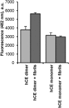
Cystatin E forms cross-β structures. A, X-ray diffraction experiments of insoluble hCE pellet revealed two rings at 10 and 4.7 Å resolution, which are characteristic for cross-β structures. B, monomeric (light gray) and dimeric (dark gray) hCE were incubated at 20, 80, and 90 °C for 10 min. Subsequently, binding of ThT was measured as an increase in fluorescence detected at 482 nm. a.u., arbitrary units. Error bars, S.D.
Figure 6.
Figure 6.
hCE fibrils contain functional protein. A, activity of legumain and papain was measured upon the addition of hCE amyloid fibrils in a fluorescent substrate assay using FR-AMC (papain) and Z-AAN-AMC (legumain) substrates. Control reactions contained only the respective enzyme. B, SDS-PAGE showing a co-precipitation assay of hCE fibrils and legumain. Insoluble hCE fibrils were incubated with active site–free legumain (AEP) and active site–blocked legumain (AEP-Dcmk). Subsequently, the insoluble fraction was harvested by centrifugation and loaded on SDS-PAGE. Control reactions contained fibrils only, AEP only, and active site–blocked legumain only. Whereas active site–free legumain bound to hCE-fibrils, active site–blocked legumain did not bind. Additionally, a band corresponding to P1-Asn39–processed hCE was observed (hCE(S40-M120); C-terminal cleavage product). Error bars, S.D.
Figure 7.
Figure 7.
hCE fibrils serve as a template for dimeric hCE to bind. hCE monomer and dimer were incubated at 90 °C for 10 min in the presence (dark gray) and absence (light gray) of hCE fibrils. Subsequently, ThT binding was measured as an increase in fluorescence at 482 nm. Control reactions contained ThT only and fibrils only. a.u., arbitrary units; Error bars, S.D.
Figure 8.
Figure 8.
hCE fibril formation is pH-dependent and incompatible with glycosylation. A, monomeric hCE was incubated at the indicated pH values for 10 min. Subsequently, binding of ThT was measured as an increase in fluorescence at 482 nm. B, glyco-hCE produced in LEXSY (Jena Bioscience) and unglycosylated hCE produced in E. coli were incubated at 90 °C for 10 min. Subsequently, binding of ThT was measured by monitoring an increase in fluorescence at 482 nm. C, model of glycosylated hCE forming a dimer but not higher oligomers. a.u., arbitrary units; Error bars, S.D.
Figure 9.
Figure 9.
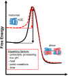
Conversion of monomeric hCE to the dimer needs a source of energy. The cystatin E monomer is a stable folding state that can convert to a dimeric state if a certain energy barrier is overcome (black arrow). The dimer has a higher thermal and fold stability as compared with the monomer. Factors reducing the energy barrier (red arrow) are time, pH, proteolytic processing, mutations, and temperature.
Figure 10.
Figure 10.
Model of cystatin E oligomerization. A, monomeric hCE can convert to a dimer upon mild conformational destabilization. Dimeric hCE can further convert to ordered oligomers potentially via concerted domain swapping reactions. The conversion from monomer to dimer and dimer to oligomer requires a certain energy barrier to be overcome. The energy barrier can be overcome by mild destabilization by low pH, N-terminal truncation, heat, and point mutations, among other factors. Presumably, dimeric hCE is a stable intermediate on the route to amyloid fibrils. Consequently, the conversion of monomeric hCE to fibrils very likely proceeds via the dimer. B, hCE fibrils are functional as legumain and papain inhibitors. For that reason, we suppose that they are heterogeneously composed of domain-swapped and monomeric subunits. The presence of hCE monomers (dark gray units) allows for the inhibition of papain-like enzymes. Inhibition of legumain is possible both with monomeric and domain-swapped hCE.

References

    1. Mason S. D., and Joyce J. A. (2011) Proteolytic networks in cancer. Trends Cell Biol. 21, 228–237 10.1016/j.tcb.2010.12.002 - DOI - PMC - PubMed
    1. Hasanbasic S., Jahic A., Karahmet E., Sejranic A., and Prnjavorac B. (2016) The role of cysteine protease in Alzheimer disease. Mater. Sociomed. 28, 235–238 10.5455/msm.2016.28.235-238 - DOI - PMC - PubMed
    1. Turk V., and Bode W. (1991) The cystatins: protein inhibitors of cysteine proteinases. FEBS Lett. 285, 213–219 10.1016/0014-5793(91)80804-C - DOI - PubMed
    1. Hedrich J., Lottaz D., Meyer K., Yiallouros I., Jahnen-Dechent W., Stöcker W., and Becker-Pauly C. (2010) Fetuin-A and cystatin C are endogenous inhibitors of human meprin metalloproteases. Biochemistry 49, 8599–8607 10.1021/bi1004238 - DOI - PubMed
    1. Abrahamson M., Barrett A. J., Salvesen G., and Grubb A. (1986) Isolation of six cysteine proteinase inhibitors from human urine: their physicochemical and enzyme kinetic properties and concentrations in biological fluids. J. Biol. Chem. 261, 11282–11289 - PubMed

Publication types

LinkOut - more resources