. 2018 Aug 22;86(9):e00239-18.
doi: 10.1128/IAI.00239-18. Print 2018 Sep.

Plasmodium vivax Merozoite Surface Protein 1 Paralog as a Mediator of Parasite Adherence to Reticulocytes

Affiliations

Plasmodium vivax Merozoite Surface Protein 1 Paralog as a Mediator of Parasite Adherence to Reticulocytes

Jin-Hee Han et al. Infect Immun. .

Abstract

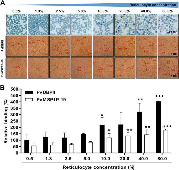
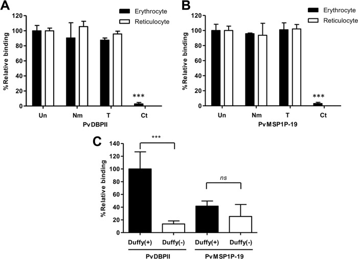
Plasmodium vivax parasites preferentially invade reticulocytes in human beings. P. vivax merozoite surface protein 1 (PvMSP1) and PvMSP1 paralog (PvMSP1P) may have important functions in reticulocyte adherence during invasion. These proteins share similar structures, including the presence of two epidermal growth factor (EGF)-like and glycosylphosphatidylinositol (GPI)-anchored domains at the C terminus. However, there have been no reports concerning the functional activity of PvMSP1P in reticulocyte adherence during P. vivax invasion. In this study, the ability of PvMSP1P-19 to bind to reticulocytes and normocytes was analyzed. The reticulocyte binding activity of PvMSP1P-19 was 4.0-fold higher than its normocyte binding activity. The binding of PvMSP1P-19 to reticulocytes and normocytes was inhibited in a dose-dependent manner by antibodies from immunized rabbits and by antibodies from vivax parasite-infected patients. Consistently, antibodies against PvMSP1P inhibited parasite invasion during short-term in vitro cultivation. Similar to the case for PvDBPII binding activity, PvMSP1P-19 binding activity was reduced in chymotrypsin-treated reticulocytes. However, no significant difference between the binding of PvMSP1P-19 to Duffy-positive and Duffy-negative erythrocytes was found. The minimal binding motif of PvMSP1P-19 was characterized using synthetic peptides. The results showed that the residues at amino acid positions 1791 to 1808 may have an important function in mediating merozoite adherence to reticulocytes. The positively charged residues within the EGF-like domain were shown to constitute a key binding motif. This work presents strong evidence supporting the role of PvMSP1P in host target cell selection and invasion of Duffy-independent pathway in P. vivax Moreover, PvMSP1P-19-specific antibodies may confer protection against P. vivax reinvasion.

Keywords: Duffy independent; Plasmodium vivax; invasion inhibition; merozoite surface; merozoite surface protein 1; merozoite surface protein 1 paralog; reticulocyte.


Figures

FIG 1
FIG 1
Schematic structure of PvMSP1P and EGF-like domain sequence homology in various malaria-causing species. (A) Schematic diagram of PvMSP1P-19 at the amino acid level. Two EGF-like domains (gray) are at aa 1751 to 1789 and 1792 to 1834. The signal peptide (SP) (black), tandem repeat region (TR) (orange), Glu/Gln polymorphic region (PR) (yellow) and GPI-anchored domain (blue) are indicated. The synthesized peptides (18-mers) are shown by the black bars within the EGF-like domain. The amino acid sequence is shown in Table S1 in the supplemental material. (B) Sequence alignment of the MSP1P-EGF-like domains of various Plasmodium species. The red bars represent sequences that are identical in all species. The sequence similarity is indicated as four (orange), three (green), or two (blue) identical species. The cysteine residue connection line represents the typical EGF-like domain disulfide bond. (C) Three-dimensional ribbon diagram of PvMSP1P-19. EGF-like domain 1 is shown in red, domain 2 is shown in gray, and cysteine residues are shown in yellow. The numbers on the cysteine residues represent the disulfide bond. (D) Three-dimensional surface diagram of PvMSP1P-19. The electrostatic surface of PvMSP1P-19 with positive (blue) and negative (red) charges is shown.
FIG 2
FIG 2
Reticulocyte binding preference. (A) Reticulocytes that were serially diluted from 80% to 0.5% were stained with new methylene blue and observed under light microscopy (upper panels). Reticulocyte binding was confirmed by PvMSP1P-19- or PvDBPII-expressed COS-7 cell rosette formation (middle and bottom panels and see Fig. S1 in the supplemental material). Red arrowheads indicate typical rosette formation. (B) The relative binding abilities at different reticulocyte concentrations were calculated and normalized to PvDBPII (100% COS-7 cell transfection efficiency) with 0.5% reticulocyte (peripheral blood condition) binding ability as a standard (100%). The data are presented as the mean ± SD of the relative binding (percent) observed in three independent experiments. PvDBPII and PvMSP1P-19 showed significant differences in binding ability under other conditions compared with the 0.5% reticulocyte condition in the PvDBPII and PvMSP1P-19 fractions, respectively. P values were calculated using Student's t test. Significant differences are indicated by single asterisks (P < 0.05), double asterisks (P < 0.01), and triple asterisks (P < 0.001).
FIG 3
FIG 3
Inhibition of erythrocyte and reticulocyte binding and P. vivax invasion ability by antibodies. (A and B) Inhibition of PvDBPII binding by antibodies against PvDBPII (A) and inhibition of PvMSP1P-19 binding by antibodies against PvMSP1P-19 (B). Upper panels, confocal microscopy images show PvDBPII and PvMSP1P-19 expression on the surface of COS-7 cells; this expression was confirmed by GFP and specific antibody recognition using antibodies against PvDBPII and antibodies against PvMSP1P-19, respectively. Lower panels, data are shown as the mean ± SD of the binding inhibition measured in three independent experiments. Significant differences in the effects of PI sera and those of other antibodies were calculated using Student's t test: single asterisks, P < 0.05; double asterisks, P < 0.01; triple asterisks, P < 0.001. (C) Inhibitory activity of serial dilutions of sera from P. vivax-infected patients (ROK+) or from uninfected persons (ROK−) on binding to erythrocytes and reticulocytes. Significant differences in the effects of ROK− and ROK+ sera were calculated using Student's t test: single asterisks, P < 0.05; double asterisks, P < 0.01; triple asterisks, P < 0.001. (D) The vivax parasite invasion inhibition efficacy was confirmed in invasion inhibition assays. The data are presented as the mean ± SD of the invasion inhibition rate obtained with preimmune sera (PI) (n = 7), anti-2C3 antibody (murine anti-Fy6) (n = 7), anti-PvMSP1-19 sera (n = 7), anti-PvMSP1P-19 sera (n = 7), and anti-PvDBPII sera (n = 5). Significant differences between PI sera and anti-2C3, anti-PvMSP1-19, anti-PvMSP1P-19, and anti-PvDBPII immunized sera were calculated using one-way ANOVA with Tukey's posttest: single asterisks, P < 0.05; double asterisks, P < 0.01; triple asterisks, P < 0.001.
FIG 4
FIG 4
Erythrocyte binding motif and residue identification. (A and B) Erythrocyte (A) and reticulocyte (B) competition binding assays were performed with PvMSP1P-19 peptides as competitors. PvDBPII was also included in an experiment to confirm nonspecific masking on RBCs. The data were analyzed using Student's t test: single asterisks, P < 0.05; double asterisks, P < 0.01. The mean ± SD of the binding inhibition rate obtained in three independent experiments is shown. (C) Inhibition of the binding of PvMSP1P-19 to erythrocytes and reticulocytes via the serial dilution of the S1764 (no inhibition detected at 100 μM) and N1791 (highest inhibition detected at 100 μM) peptides. The data are presented as the mean ± SD of the binding inhibition observed in three independent experiments. (D) Reticulocyte binding ability of mutant PvMSP1P-19 protein. The data are presented as the mean ± SD of the percentage of relative binding of PvMSP1P-19 observed in three independent experiments using the Sal-1 strain. Significant differences were calculated using one-way ANOVA with Tukey's posttest: single asterisks, P < 0.05; double asterisks, P < 0.01. (E) Sites of amino acid mutation are highlighted in the sequence alignment. The blue box shows the position of an unimportant peptide (S1764). The red box represents an important region for reticulocyte binding as confirmed by peptide competition assays. The red star indicates a critical residue for reticulocyte binding. (F) Three-dimensional structure of the reticulocyte binding region of PvMSP1P-19.
FIG 5
FIG 5
Reticulocyte-specific receptors for PvMSP1P-19 binding. (A and B) PvDBPII (A) and PvMSP1P-19 (B) binding assays were performed using enzyme-treated erythrocytes and reticulocytes. Untreated (Un) erythrocytes and reticulocytes and erythrocytes and reticulocytes treated with neuraminidase (Nm), trypsin (T), or chymotrypsin (Ct) were used to confirm receptor specificity. The data are shown as the mean ± SD of the binding inhibition rate measured in three independent experiments. Significant differences compared to the results with untreated erythrocytes or reticulocytes after the enzymatic treatment of erythrocytes and reticulocytes were calculated using Student's t test and are denoted by triple asterisks (P < 0.001). (C) Binding specificity of PvMSP1P-19 and PvDBPII to Duffy-positive and Duffy-negative erythrocytes. The relative binding of PvMSP1P-19 to Duffy-positive and Duffy-negative erythrocytes was normalized to that of PvDBPII binding to Duffy-positive erythrocytes as a standard (100%). The data are shown as the mean ± SD of the relative binding measured in four independent experiments. Significant differences between Duffy-positive and Duffy-negative erythrocytes in PvDBPII and PvMSP1P-19, respectively, were observed. P values were calculated using Student's t test; significant differences are indicated by triple asterisks (P < 0.001). ns, not significant.

References

    1. World Health Organization. 2017. World malaria report 2016. World Health Organization, Geneva, Switzerland.
    1. Lim C, Pereira L, Saliba KS, Mascarenhas A, Maki JN, Chery L, Gomes E, Rathod PK, Duraisingh MT. 2016. Reticulocyte preference and stage development of Plasmodium vivax isolates. J Infect Dis 214:1081–1084. doi:10.1093/infdis/jiw303. - DOI - PMC - PubMed
    1. Chitnis CE, Sharma A. 2008. Targeting the Plasmodium vivax Duffy-binding protein. Trends Parasitol 24:29–34. doi:10.1016/j.pt.2007.10.004. - DOI - PubMed
    1. Malleret B, Xu F, Mohandas N, Suwanarusk R, Chu C, Leite JA, Low K, Turner C, Sriprawat K, Zhang R, Bertrand O, Colin Y, Costa FT, Ong CN, Ng ML, Lim CT, Nosten F, Renia L, Russell B. 2013. Significant biochemical, biophysical and metabolic diversity in circulating human cord blood reticulocytes. PLoS One 8:e76062. doi:10.1371/journal.pone.0076062. - DOI - PMC - PubMed
    1. Menard D, Barnadas C, Bouchier C, Henry-Halldin C, Gray LR, Ratsimbasoa A, Thonier V, Carod JF, Domarle O, Colin Y, Bertrand O, Picot J, King CL, Grimberg BT, Mercereau-Puijalon O, Zimmerman PA. 2010. Plasmodium vivax clinical malaria is commonly observed in Duffy-negative Malagasy people. Proc Natl Acad Sci U S A 107:5967–5971. doi:10.1073/pnas.0912496107. - DOI - PMC - PubMed

Publication types

LinkOut - more resources