. 2018 Aug 22;86(9):e00322-18.
doi: 10.1128/IAI.00322-18. Print 2018 Sep.

Gene Regulation by Redox-Sensitive Burkholderia thailandensis OhrR and Its Role in Bacterial Killing of Caenorhabditis elegans

Affiliations

Gene Regulation by Redox-Sensitive Burkholderia thailandensis OhrR and Its Role in Bacterial Killing of Caenorhabditis elegans

Anuja Pande et al. Infect Immun. .

Abstract

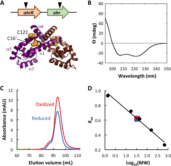
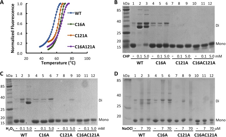
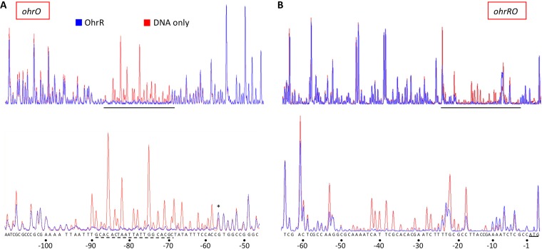
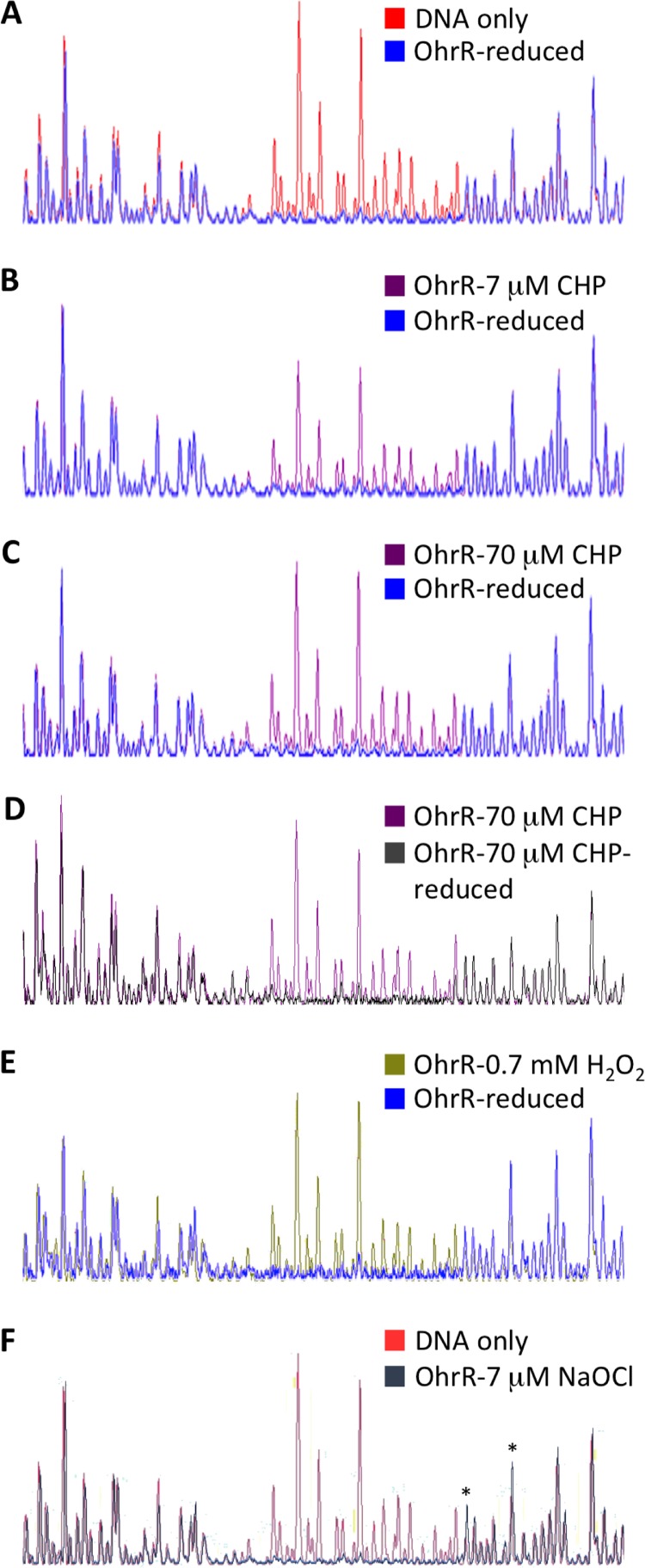
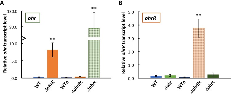
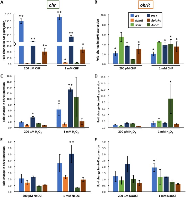
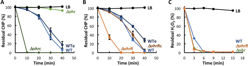
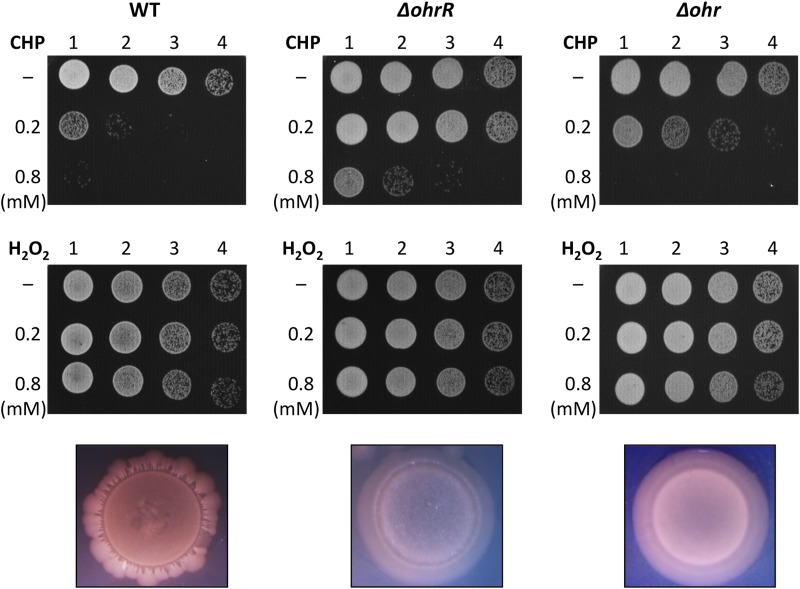
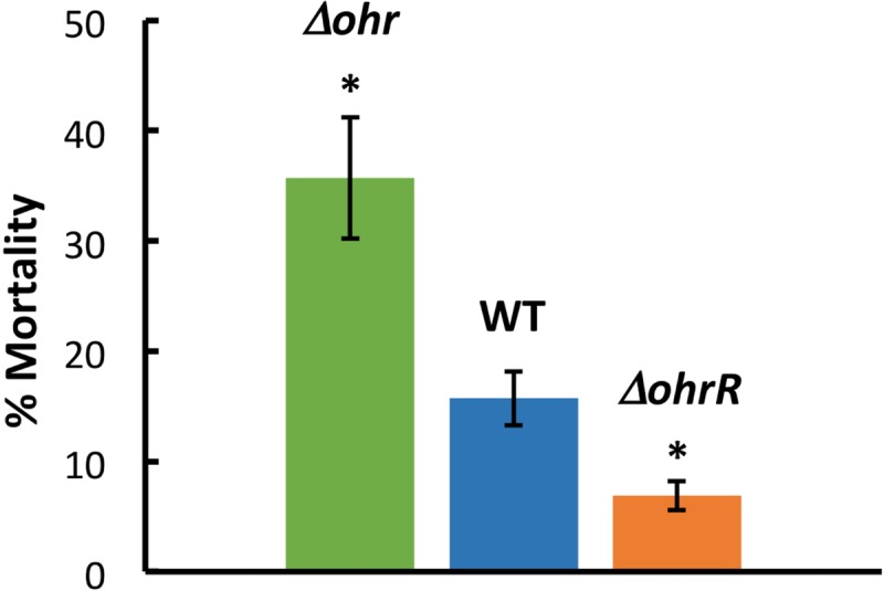
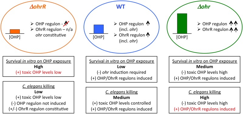
Fatty acid hydroperoxides are involved in host-pathogen interactions. In both plants and mammals, polyunsaturated fatty acids are liberated during infection and enzymatically oxidized to the corresponding toxic hydroperoxides during the defensive oxidative burst that is designed to thwart the infection. The bacterial transcription factor OhrR (organic hydroperoxide reductase regulator) is oxidized by organic hydroperoxides, as a result of which the ohr gene encoding organic hydroperoxide reductase is induced. This enzyme converts the hydroperoxides to less toxic alcohols. We show here that OhrR from Burkholderia thailandensis represses expression of ohr Gene expression is induced by cumene hydroperoxide and to a lesser extent by inorganic oxidants; however, Ohr contributes to degradation only of the organic hydroperoxide. B. thailandensis OhrR, which binds specific sites in both ohr and ohrR promoters, as evidenced by DNase I footprinting, belongs to the 2-Cys subfamily of OhrR proteins, and its oxidation leads to reversible disulfide bond formation between conserved N- and C-terminal cysteines in separate monomers. Oxidation of the N-terminal Cys is sufficient for attenuation of DNA binding in vitro, with complete restoration of DNA binding occurring on addition of a reducing agent. Surprisingly, both overexpression of ohr and deletion of ohr results in enhanced survival on exposure to organic hydroperoxide in vitro While Δohr cells are more virulent in a Caenorhabditis elegans model of infection, ΔohrR cells are less so. Taken together, our data suggest that B. thailandensis OhrR has several unconventional features and that both OhrR and organic hydroperoxides may contribute to virulence.

Keywords: DNase I footprinting; MarR; Ohr; ROS; gene regulation; organic hydroperoxide; transcription factors.


Figures

FIG 1
FIG 1
B. thailandensis OhrR is a dimer. (A) (Top) The genomic locus encoding ohr and ohrR. The inverted triangles indicate the positions of transposon insertion. (Bottom) The model is based on the structure of reduced X. campestris OhrR-C22S (PDB accession number 2PEX [19]; global model quality estimate [GMQE] = 0.72). One subunit is in magenta with helices identified, and the other is in brown. Cysteine residues are in yellow space-filling representation, with C16′ in α1′ (brown subunit) and C121 in α5 (magenta subunit) being identified. The illustration was generated with the PyMOL program. (B) Far-UV CD spectrum reflecting the expected secondary structure composition. Ellipticity (θ) is reported in millidegrees (mdeg). (C) Elution of reduced and oxidized OhrR from SEC column. mAU, milli-absorbance units. (D) Standard curve reflecting MW standards eluting from the SEC column. Both reduced (blue) and oxidized (red) OhrR eluted corresponding to an MW of ∼34 kDa.
FIG 2
FIG 2
In vitro oxidation and stability of OhrR variants. (A) Thermal stability of OhrR variants determined by fluorescence of SYPRO Orange bound to unfolded protein as a function of temperature. (B to D) Oxidant-treated OhrR variants separated on 12% nonreducing SDS-PAGE gels. Migration corresponding to the monomer and dimer, identified at the right. Lanes marked kDa, MW markers; lanes 1 to 3, WT OhrR; lanes 4 to 6, OhrR-C16A; lanes 7 to 9, OhrR-C121A; lanes 10 to 12, OhrR-C16AC121A. The addition of CHP (0.1 or 5.0 mM; B), H2O2 (0.1 or 5.0 mM; C), or NaOCl (7 or 70 μM; D) is identified below each gel.
FIG 3
FIG 3
OhrR binds specifically to ohr and ohrR promoter DNA. (A) Electropherogram traces of DNase I-digested ohr promoter DNA. (B) Electropherogram traces of DNase I-digested ohrR promoter DNA. Red, DNA only; blue, DNA incubated with OhrR at a stoichiometric DNA/protein ratio of 1:4. The protected regions are expanded in the lower panels, with the sequence being identified at the bottom. Numbering is relative to the translational start, defined as position +1. For ohrR promoter DNA, the ATG codon is underlined. For ohr promoter DNA, a sequence resembling the published OhrR consensus sequence is identified by a dashed underline; the asterisk marks a hypersensitive site. The results are representative of those from at least three replicates.
FIG 4
FIG 4
Effect of OhrR oxidation on binding to ohr promoter DNA. (A) Electropherogram traces of DNase I-digested ohr promoter DNA and DNA incubated with reduced OhrR. (B) DNA incubated with reduced OhrR or with OhrR oxidized with 7 μM CHP. (C) DNA incubated with reduced OhrR or with OhrR oxidized with 70 μM CHP. (D) DNA incubated with OhrR oxidized with 70 μM CHP or with 70 μM CHP-oxidized OhrR subsequently rereduced with DTT. (E) DNA incubated with reduced OhrR or with OhrR oxidized with 0.7 mM H2O2. (F) Free ohr promoter DNA and DNA incubated with OhrR oxidized with 7 μM NaOCl. Asterisks mark hypersensitive sites (positions −56 and −49). The results are representative of those from at least three replicates.
FIG 5
FIG 5
OhrR represses ohr expression. (A) Expression of ohr in WT and WTe (the WT strain containing the empty plasmid) cells and in ΔohrR and ΔohrRc (the ΔohrR strain complemented with ohrR) cells. The hatched green bar confirms abundant ohr expression in Δohr cells complemented with ohrohrc). (B) Expression of ohrR in WT and WTe cells and in Δohr and Δohrc (the Δohr strain complemented with ohr) cells. The hatched orange bar confirms abundant ohrR expression in ΔohrR cells complemented with ohrR. Transcript levels are reported relative to the level of transcription of the reference gene (glutamate synthase) and were determined using the 2−ΔCT method. Error bars represent standard errors from three independent experiments. Asterisks directly above the bars denote statistically significant differences in expression compared to that in WT cells, as determined by an unpaired Student t test (**, P < 0.001).
FIG 6
FIG 6
Gene expression is induced by oxidants. (A) Fold change in ohr expression after incubation of cells with 0.2 or 1 mM CHP relative to that in unsupplemented cultures. (B) Fold change in ohrR expression after incubation of cells with 0.2 or 1 mM CHP. (C, E) Fold change in ohr expression after incubation of cells with 0.2 or 1 mM H2O2 (C) or NaOCl (E) relative to that in unsupplemented cultures. (D, F) Fold change in ohrR expression after incubation of cells with 0.2 or 1 mM H2O2 (D) or NaOCl (F). Color coding is defined in the inset to panel B and is identical for all panels. Transcript levels were reported using the 2−ΔΔCT method. Error bars represent standard errors from at least two independent experiments. Asterisks directly above the bars denote a statistically significant difference in expression compared to that in the corresponding unsupplemented culture, as determined by an unpaired Student t test (*, P < 0.05; **, P < 0.001).
FIG 7
FIG 7
Determination of residual oxidant in cell culture medium. The relative levels of oxidants added at 0 min were determined using the FOX assay. (A, B) 200 μM CHP; (C) 200 μM H2O2. Oxidant-containing LB medium was used as a control. Where error bars (representing the SD from three experiments) are missing, they are smaller than the symbol sizes.
FIG 8
FIG 8
Effect of ohr and ohrR deletions on survival and colony morphology. WT, ΔohrR, and Δohr cells were exposed to the identified concentrations of CHP (top row) or H2O2 (middle row) for 15 min, following which 10-fold serial dilutions were spotted on agar plates. The results are representative of those from at least three experiments. The corresponding colony morphologies, captured after ∼72 h, are illustrated at the bottom.
FIG 9
FIG 9
Pathogenicity of B. thailandensis in C. elegans. Percentage of dead worms 12 h after depositing worms (L4 larval stage) on plates inoculated with the identified B. thailandensis strain. Asterisks denote a P value of <0.05 compared to the WT, based on an unpaired t test. Data are from three independent experiments, with error bars indicating standard errors.
FIG 10
FIG 10
Deletion of ohr or ohrR differentially affects survival on exposure to organic hydroperoxides in vitro and killing of C. elegans. Ovals represent ΔohrR, WT, and Δohr cells in which the levels of organic hydroperoxide (OHP) are low, medium, and high, respectively (colored bar graphs), as determined by the FOX assay (Fig. 7). Genes in the OHP and/or OhrR regulons (including ohr, in the case of WT cells) are expected to be the most efficiently induced in WT and Δohr cells. Boxes below the ovals represent items that may promote (+) or counter (−) survival on exposure to OHP in vitro and C. elegans killing. n/a, not applicable.

Similar articles

Cited by

References

    1. Sies H, Berndt C, Jones DP. 2017. Oxidative stress. Annu Rev Biochem 86:715–748. doi:10.1146/annurev-biochem-061516-045037. - DOI - PubMed
    1. Knoefler D, Leichert LI, Thamsen M, Cremers CM, Reichmann D, Gray MJ, Wholey WY, Jakob U. 2014. About the dangers, costs and benefits of living an aerobic lifestyle. Biochem Soc Trans 42:917–921. doi:10.1042/BST20140108. - DOI - PMC - PubMed
    1. Rosen H, Crowley JR, Heinecke JW. 2002. Human neutrophils use the myeloperoxidase-hydrogen peroxide-chloride system to chlorinate but not nitrate bacterial proteins during phagocytosis. J Biol Chem 277:30463–30468. doi:10.1074/jbc.M202331200. - DOI - PubMed
    1. Nathan C, Ding A. 2010. SnapShot: reactive oxygen intermediates (ROI). Cell 140:951–951.e952. doi:10.1016/j.cell.2010.03.008. - DOI - PubMed
    1. Klomsiri C, Panmanee W, Dharmsthiti S, Vattanaviboon P, Mongkolsuk S. 2005. Novel roles of ohrR-ohr in Xanthomonas sensing, metabolism, and physiological adaptive response to lipid hydroperoxide. J Bacteriol 187:3277–3281. doi:10.1128/JB.187.9.3277-3281.2005. - DOI - PMC - PubMed

Publication types

MeSH terms

LinkOut - more resources