. 2018 Jul 17;115(29):E6731-E6740.
doi: 10.1073/pnas.1721431115. Epub 2018 Jul 2.

Decoding on the ribosome depends on the structure of the mRNA phosphodiester backbone

Affiliations

Decoding on the ribosome depends on the structure of the mRNA phosphodiester backbone

Hannah E Keedy et al. Proc Natl Acad Sci U S A. .

Abstract

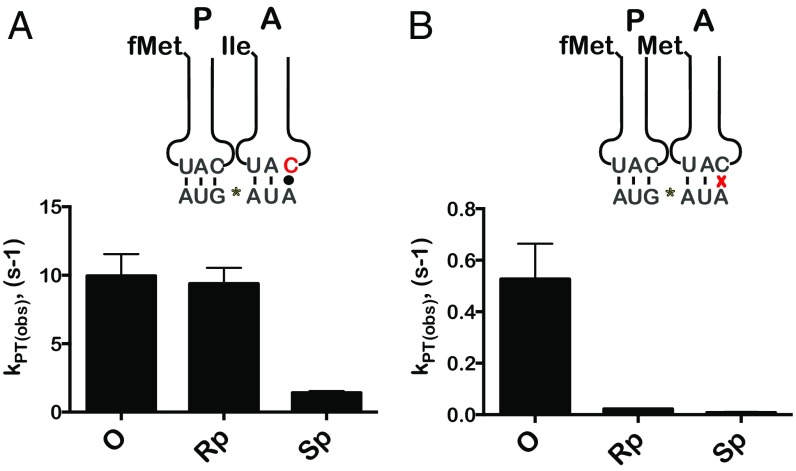
During translation, the ribosome plays an active role in ensuring that mRNA is decoded accurately and rapidly. Recently, biochemical studies have also implicated certain accessory factors in maintaining decoding accuracy. However, it is currently unclear whether the mRNA itself plays an active role in the process beyond its ability to base pair with the tRNA. Structural studies revealed that the mRNA kinks at the interface of the P and A sites. A magnesium ion appears to stabilize this structure through electrostatic interactions with the phosphodiester backbone of the mRNA. Here we examined the role of the kink structure on decoding using a well-defined in vitro translation system. Disruption of the kink structure through site-specific phosphorothioate modification resulted in an acute hyperaccurate phenotype. We measured rates of peptidyl transfer for near-cognate tRNAs that were severely diminished and in some instances were almost 100-fold slower than unmodified mRNAs. In contrast to peptidyl transfer, the modifications had little effect on GTP hydrolysis by elongation factor thermal unstable (EF-Tu), suggesting that only the proofreading phase of tRNA selection depends critically on the kink structure. Although the modifications appear to have no effect on typical cognate interactions, peptidyl transfer for a tRNA that uses atypical base pairing is compromised. These observations suggest that the kink structure is important for decoding in the absence of Watson-Crick or G-U wobble base pairing at the third position. Our findings provide evidence for a previously unappreciated role for the mRNA backbone in ensuring uniform decoding of the genetic code.

Keywords: decoding; mRNA structure; phosphorothioate substitution; ribosome; tRNA selection.


Conflict of interest statement

The authors declare no conflict of interest.

Figures

Fig. 1.
Fig. 1.
Structure of the mRNA on the ribosome and preparation of phosphorothioate-modified mRNAs. (A) Overview of the mRNA structure [Protein Data Bank (PDB) ID code 2J00] highlighting the kink structure dividing the P and A sites of the ribosome. The nonbridging oxygen atoms (red) coordinating a magnesium ion (green) are shown. (B) A representative HPLC chromatogram showing the separation of the two phosphorothioate diastereoisomers of the downstream RNA sequence. (C) Schematic of the procedure used to synthesize the full-length modified mRNA. (Upper) Sequences of the two pieces are shown annealed to a DNA splint. (Lower) A representative denaturing PAGE used to follow the ligation reaction using a radiolabeled downstream sequence.
Fig. 2.
Fig. 2.
Phosphorothioate mRNAs suppress the incorporation of near-cognate amino acids PhosphorImager scans of electrophoretic TLCs showing the reactivity profile of the initiation complexes programmed with the indicated native and phosphorothioate-modified mRNAs with the 20 aa-tRNA isoacceptors. Differential reactivities with near-cognate aa-tRNA are marked by asterisks. Note that for this particular reactivity survey the formylation of fMet was incomplete, and as a result residual Met is observed. This does not affect the analysis because of differences in migration on the TLC between fMet and Met as well as the corresponding dipeptides. A schematic of the initiation complex with fMet-tRNAfMet occupying the P site and the Glu GAA codon occupying the A siteis shown above the scans.
Fig. 3.
Fig. 3.
The Sp-phosphorothioate substitution of the kink oxygen results in a severe hyperaccurate phenotype. (A) Representative time courses for PT reactions in the indicated complexes and the cognate Glu-tRNAGlu ternary complex. (B) Representative time course for PT reactions in the indicated complexes and the near-cognate Lys-tRNALys ternary complex. (C) Representative time courses for PT reactions in the indicated complexes and the near-cognate Asp-tRNAAsp ternary complex. (D) Bar graph showing the observed rate of GTP hydrolysis for modified and Rp- and Sp- complexes with the near-cognate Lys-tRNALys ternary complex. Unlike PT reactions, which were all conducted at 37 °C, these reactions were conducted at 20 °C. Shown are the means of three independent time courses; error bars represent the SD from the mean. (E) Representative time courses for RF2-mediated hydrolysis on the indicated complexes. (F) Representative time courses for RF1-mediated hydrolysis reaction on the indicated complexes.
Fig. 4.
Fig. 4.
Substitution of the proS oxygen reduces the rate of peptide-bond formation in the presence of atypical tRNA–mRNA interactions. (A) Bar graph showing the PT rate for unmodified and Rp- and Sp- complexes, all displaying the Ile AUA codon in the A site (depicted above), with the cognate Ile-tRNAIle. The corresponding Ile-tRNA harbors the lysidine (k2C) modification at the wobble position. (B) Observed PT rates for the near-cognate Met-tRNAMet in the presence of the indicated complexes. Graphs show the means of three independent time courses; error bars represent the SD from the mean.
Fig. 5.
Fig. 5.
Peptide release is not impacted by phosphorothioate modification at interface of the P-site and A-site codons. (A and B) Representative time courses for peptide release between the indicated initiation complexes (programmed with either a native mRNA or a racemic mixture of phosphorothioate mRNAs) and RF1 and RF2, respectively.
Fig. 6.
Fig. 6.
Phosphorothioate modification at the second position of the A-site codon results in a hyperaccurate phenotype. (A) Representative time courses of peptide-bond formation between the depicted unmodified or modified complexes with the cognate Glu-tRNAGlu. The phosphorothioate modification is between the G and A of the A-site GAA codon (as shown above). (B and C) Time courses of PT reactions with the near-cognate Lys-tRNALys (B) and Asp-tRNAAsp (C) ternary complexes, respectively. (D and E) Time courses for RF1- and RF2-mediated hydrolysis reactions, respectively.
Fig. 7.
Fig. 7.
Phosphorothioate substitution between the second and third nucleotide of the A-site codon does not significantly impact PT. (A and B) Representative time courses for PT between the indicated initiation complexes and the cognate Glu-tRNAGlu (A) and near-cognate Lys-tRNALys (B). The phosphorothioate modification is between the second and third nucleotide of the A-site codon (as shown above).
Fig. 8.
Fig. 8.
Deoxyribose substitutions in the A-site codon result in a severe hyperaccurate phenotype. (A) Representative time courses for PT reactions between the initiation complexes programmed with the indicated native and deoxyribose-modified mRNAs and the cognate Glu-tRNAGlu ternary complex. (B) Representative time courses for PT reactions between the indicated complexes and the near-cognate Lys-tRNALys ternary complex. (C) Representative time course for PT between complexes displaying the native AUA codon or the deoxy-modified one at the third position of the codon (AUdA) and the cognate Ile-tRNAIle ternary complex. The corresponding Ile-tRNA harbors the lysidine (k2C) modification at the wobble position.

References

    1. Rosenberger RF, Foskett G. An estimate of the frequency of in vivo transcriptional errors at a nonsense codon in Escherichia coli. Mol Gen Genet. 1981;183:561–563. - PubMed
    1. Edelmann P, Gallant J. Mistranslation in E. coli. Cell. 1977;10:131–137. - PubMed
    1. Bouadloun F, Donner D, Kurland CG. Codon-specific missense errors in vivo. EMBO J. 1983;2:1351–1356. - PMC - PubMed
    1. Kramer EB, Farabaugh PJ. The frequency of translational misreading errors in E. coli is largely determined by tRNA competition. RNA. 2007;13:87–96. - PMC - PubMed
    1. Thompson RC. EFTu provides an internal kinetic standard for translational accuracy. Trends Biochem Sci. 1988;13:91–93. - PubMed

Publication types

MeSH terms