. 2018 Jul 23;145(14):dev165456.
doi: 10.1242/dev.165456.

SP8 and SP9 coordinately promote D2-type medium spiny neuron production by activating Six3 expression

Affiliations

SP8 and SP9 coordinately promote D2-type medium spiny neuron production by activating Six3 expression

Zhejun Xu et al. Development. .

Abstract

Dopamine receptor DRD1-expressing medium spiny neurons (D1 MSNs) and dopamine receptor DRD2-expressing medium spiny neurons (D2 MSNs) are the principal projection neurons in the striatum, which is divided into dorsal striatum (caudate nucleus and putamen) and ventral striatum (nucleus accumbens and olfactory tubercle). Progenitors of these neurons arise in the lateral ganglionic eminence (LGE). Using conditional deletion, we show that mice lacking the transcription factor genes Sp8 and Sp9 lose virtually all D2 MSNs as a result of reduced neurogenesis in the LGE, whereas D1 MSNs are largely unaffected. SP8 and SP9 together drive expression of the transcription factor Six3 in a spatially restricted domain of the LGE subventricular zone. Conditional deletion of Six3 also prevents the formation of most D2 MSNs, phenocopying the Sp8/9 mutants. Finally, ChIP-Seq reveals that SP9 directly binds to the promoter and a putative enhancer of Six3 Thus, this study defines components of a transcription pathway in a regionally restricted LGE progenitor domain that selectively drives the generation of D2 MSNs.

Keywords: DRD2; LGE; Medium spiny neuron; Mouse; Six3; Sp8; Sp9; Striatum.


Conflict of interest statement

Competing interestsJ.L.R. is a co-founder and stockholder, and is currently on the scientific board, of Neurona, a company studying the potential therapeutic use of interneuron transplantation.

Figures

Fig. 1.
Fig. 1.
Most SP8+ cells in the LGE SVZ express SP9. (A-H) Co-expression of SP8 and SP9 in the LGE SVZ at E14.5 and E16.5. Note that SP8 is strongly expressed in the dLGE SVZ, and weakly expressed in the vLGE SVZ, but most SP8+ cells express SP9. (I,J) SP8 expression in the E16.5 LGE SVZ (rostral to the left) of wild-type and Sp9-KO mice. Arrows indicate upregulation of SP8 in the LGE. Dotted line indicates the border of the dorsal and ventral LGE. Scale bars: 100 µm in G for A-C,E-G; 20 µm in H for D,H; 200 µm in J for I,J.
Fig. 2.
Fig. 2.
The Sp9-KO striatum has more SP8+/DRD2-GFP+ MSNs. (A-D) SP8/GFP double immunostained coronal striatal sections of Drd2-GFP control and Drd2-GFP; Sp9lacZ/lacZ (Sp9-KO) mutant mice at P9. (E-L) Higher magnification images of the boxed areas in A-D. (M,N) Quantification data showing that Sp9-KO mice had more SP8+ and SP8+/DRD2-GFP+ cells in the striatum per section than controls (Student's t-test, *P<0.05, **P<0.01, n=3, mean+s.e.m.). CPu, caudate-putamen; NAc, nucleus accumbens; OT, olfactory tubercle. Scale bars: 200 µm in D for A-D; 50 µm in L for E-L.
Fig. 3.
Fig. 3.
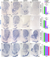
Sp8/9-DCKO mice lose nearly all D2 MSNs. (A-J) In situ RNA hybridization of Drd2 and Adora2a showed that nearly all D2 MSNs were lost in the striatum at P11. Those remaining Drd2+ cells in the Sp8/9-DCKO striatum (D) are ChAT+ interneurons. (K-T) Drd1 and Tac1 mRNA in the striatum of control and mutant mice at P11. The generation of D1 MSNs was largely unaffected in mutant mice relative to controls. (E,J,O,T) Histograms showing quantification of the data (one-way ANOVA followed by Tukey–Kramer post-hoc test, **P<0.01, ***P<0.001, n=3, mean+s.e.m.). Scale bar: 500 µm in S for A-D,F-I,K-N,P-S.
Fig. 4.
Fig. 4.
Accumulation of neural progenitors in the LGE of Sp8/9-DCKO mice at E16.5. (A-D) In situ RNA hybridization showed that there were more neural stem/progenitor cells (Gsx2+, Dlx1+, Dlx2+ and Ascl1+ cells, arrows) in the LGE VZ/SVZ of Sp8-CKO and Sp8/9-DCKO mice compared with control and Sp9-KO mice. (E-L′) GSX2 and ASCL1 immunostained LGE sections in control and mutant mice. Dotted lines mark the border of the VZ and SVZ of the LGE. (M-P) Quantification data showing that there are more GSX2+ and ASCL1+ neural progenitors in the LGE VZ and SVZ of Sp8-CKO and Sp8/9-DCKO mice. (one-way ANOVA followed by Tukey–Kramer post-hoc test, **P<0.01, ***P<0.001, n=3, mean+s.e.m.). Scale bars: 200 µm in D for A-D; 200 µm in L for E-L; 50 µm in L′ for E′-L′.
Fig. 5.
Fig. 5.
MSN production is reduced in Sp8/9-DCKO mice. (A-J) The Sp8-CKO, Sp9-KO and Sp8/9-DCKO LGE SVZ had fewer FOXP1+ and EBF1+ cells relative to wild-type controls at E16.5. (K-N) DCX and TUBB3 expression was greatly reduced in Sp8/9-DCKO LGE SVZ. (O-T) BrdU was injected intraperitoneally at E15.5, and mice were sacrificed at E16.5 (24 h after BrdU injection). Sp8-CKO and Sp8/9-DCKO embryos had more BrdU+ cells in the LGE VZ/SVZ relative to controls, whereas Sp9-KO and Sp8/9-DCKO embryos had more BrdU+ cells in the LGE MZ (striatum). (U-Y) BrdU was injected intraperitoneally at E13.5, and then mice were sacrificed at P0. Immunostaining of BrdU showed that mutant mice had reduced BrdU+ cells in the striatum relative to controls. Dotted lines mark the border of the VZ, SVZ and MZ of the LGE. CPu, caudate-putamen. One-way ANOVA followed by Tukey–Kramer post-hoc test, *P<0.05, **P<0.01, ***P<0.001, n=3, mean+s.e.m.). Scale bars: 100 µm in S for A-D,F-I,P-S; in X for U-X.
Fig. 6.
Fig. 6.
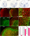
Six3 expression in the LGE. (A-C) In situ hybridization shows Six3 expression in the ganglionic eminences, weak Six3 expression in the VZ and strong Six3 expression in the dorsal part of the vLGE SVZ. (D-F) Immunostaining of BrdU and SIX3 on LGE sections at E16.5, 30 min after a BrdU pulse. Arrows point to BrdU+/SIX3+ cells. Note that SIX3 expression was downregulated from the SVZ to the striatum. (G-J) Immunostaining of DRD2-GFP, SIX3 and SP9 on LGE sections at E16.5. The dLGE SVZ did not express SIX3. (K-M) Quantification of immunostaining experiments showing that, in the LGE SVZ, most DRD2-GFP+ cells expressed SIX3 and most SIX3+ cells expressed SP9. n=3, mean+s.e.m. Scale bars: 200 µm in C for A-C; 200 µm in D for D,E; 100 µm in H for G,H; 50 µm in I for F,I; 50 µm in J.
Fig. 7.
Fig. 7.
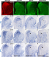
Sp8/9-DCKO mice fail to express Six3 in the LGE SVZ. (A-D) Immunostaining images show expressions of SP9, Dlx5/6-GFP, SP9-lacZ and Dlx5/6-GFP in the LGE of wild-type, Sp8-CKO, Sp9-KO and Sp8/9-DCKO mice, respectively. (E-L) In situ hybridization showing Six3 expression in wild-type and mutant LGE at E14.5 and E16.5. Six3 expression was reduced in the Sp9-KO LGE SVZ (arrowheads), and was almost undetectable in the Sp8/9-DCKO LGE SVZ (arrows), whereas Six3 expression in the LGE of Sp8-CKO mice was less affected. (M-P) Six3 expression in the LGE SVZ was also undetectable (P) when a Nestin-Cre transgenic allele was used to knockout Sp8 and Sp9. Scale bar: 200 µm in P for A-P.
Fig. 8.
Fig. 8.
Most D2 MSNs were lost in the striatum of Six3-CKO mice. (A-L) In situ RNA hybridization of Drd2, Adora2a, Penk and Gpr6 showing that >90% of D2 MSNs were lost in the striatum of Six3-CKO mice compared with controls (Dlx5/6-CIE) at P11. (M-R) The generation of D1 MSNs (Drd1+ and Tac1+ cells) was less affected. Note that the lateral ventricle was enlarged in Six3-CKO mice. (Student's t-test, **P<0.01, ***P<0.001, n=3, mean+s.e.m.). Scale bar: 500 µm in Q for A-Q.
Fig. 9.
Fig. 9.
SP9 binds to the promoter of Six3. (A) Venn diagram showing overlap of called peaks between SP9 ChIP at E13.5 and E16.5. (B) Pie charts showing genomic distribution of SP9-binding peaks (for E13.5 and E16.5 experiments) relative to promoters (within ±1 kb of TSS), 5′ UTRs, exons, introns, 3′ UTRs and intergenic regions. (C) ChIP-Seq genome browser tracks from E13.5 (green) and E16.5 (black) experiments, showing SP9 ChIP, input and ChIP versus input. Small rectangles beneath the tracks indicate called peaks. Large red rectangles highlight where SP9 binds to the promoter and several upstream regions (regions 1, 2 and 3) of the Six3 gene. (D) SP9 consensus motif and relative enrichment derived from de novo motif analysis of intersected SP9 ChIP-Seq. (E) Top three frequent motifs (according to P-value) derived from the intersection of two ChIP experiments, displaying the associated transcription factor and percentage of total peaks. (F) SP9-activated transcription from the Six3 promoter and a putative enhancer in a dual-luciferase assay within P19 cells. P, promoter; R1, region 1; R2, region 2; R3, region 3. Student's t-test, *P<0.05, **P<0.01, n=3 individuals, mean+s.e.m.

Similar articles

Cited by

References

    1. Appolloni I., Calzolari F., Corte G., Perris R. and Malatesta P. (2008). Six3 controls the neural progenitor status in the murine CNS. Cereb. Cortex 18, 553-562. 10.1093/cercor/bhm092 - DOI - PubMed
    1. Bell S. M., Schreiner C. M., Waclaw R. R., Campbell K., Potter S. S. and Scott W. J. (2003). Sp8 is crucial for limb outgrowth and neuropore closure. Proc. Natl. Acad. Sci. USA 100, 12195-12200. 10.1073/pnas.2134310100 - DOI - PMC - PubMed
    1. Castro D. S., Martynoga B., Parras C., Ramesh V., Pacary E., Johnston C., Drechsel D., Lebel-Potter M., Garcia L. G., Hunt C. et al. (2011). A novel function of the proneural factor Ascl1 in progenitor proliferation identified by genome-wide characterization of its targets. Genes Dev. 25, 930-945. 10.1101/gad.627811 - DOI - PMC - PubMed
    1. Del Bene F. and Wittbrodt J. (2005). Cell cycle control by homeobox genes in development and disease. Semin. Cell Dev. Biol. 16, 449-460. 10.1016/j.semcdb.2005.02.001 - DOI - PubMed
    1. Del Bene F., Tessmar-Raible K. and Wittbrodt J. (2004). Direct interaction of geminin and Six3 in eye development. Nature 427, 745-749. 10.1038/nature02292 - DOI - PubMed

Publication types

MeSH terms