. 2018 Jul 2;9(1):2563.
doi: 10.1038/s41467-018-05004-y.

BrlR from Pseudomonas aeruginosa is a receptor for both cyclic di-GMP and pyocyanin

Affiliations

BrlR from Pseudomonas aeruginosa is a receptor for both cyclic di-GMP and pyocyanin

Feng Wang et al. Nat Commun. .

Abstract

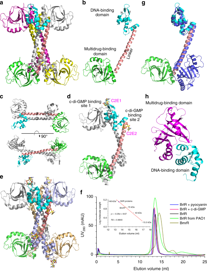
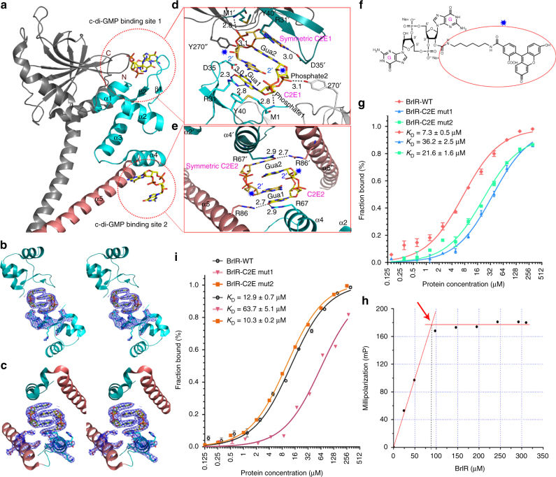
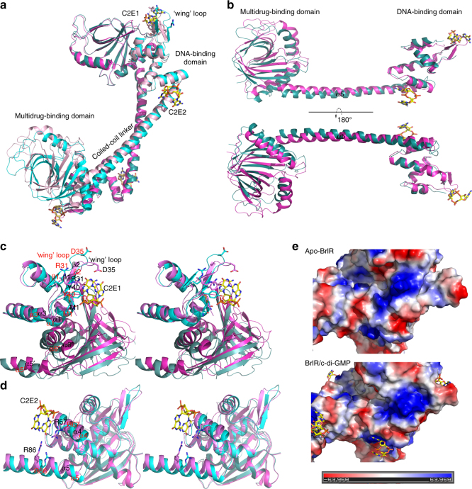
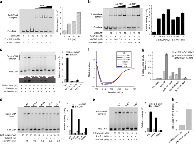
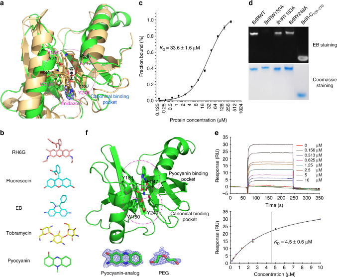
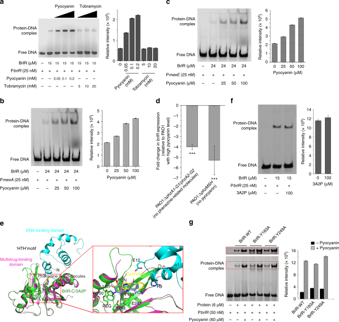
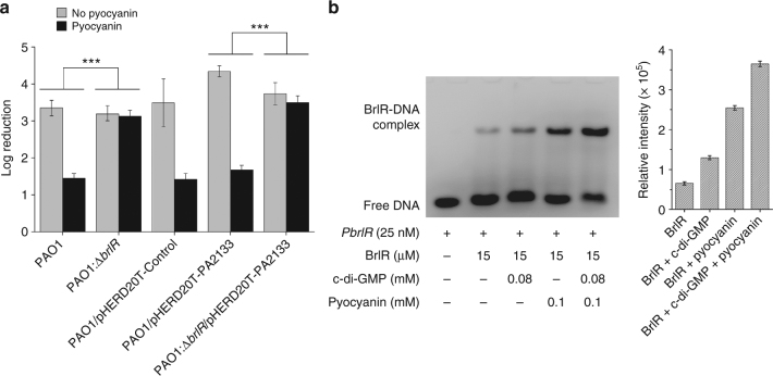
The virulence factor pyocyanin and the intracellular second messenger cyclic diguanylate monophosphate (c-di-GMP) play key roles in regulating biofilm formation and multi-drug efflux pump expression in Pseudomonas aeruginosa. However, the crosstalk between these two signaling pathways remains unclear. Here we show that BrlR (PA4878), previously identified as a c-di-GMP responsive transcriptional regulator, acts also as a receptor for pyocyanin. Crystal structures of free BrlR and c-di-GMP-bound BrlR reveal that the DNA-binding domain of BrlR contains two separate c-di-GMP binding sites, both of which are involved in promoting brlR expression. In addition, we identify a pyocyanin-binding site on the C-terminal multidrug-binding domain based on the structure of the BrlR-C domain in complex with a pyocyanin analog. Biochemical analysis indicates that pyocyanin enhances BrlR-DNA binding and brlR expression in a concentration-dependent manner.


Conflict of interest statement

The authors declare no competing interests.

Figures

Fig. 1
Fig. 1
Crystal structures of BrlR and the BrlR–c-di-GMP complex. a Overall structure of the apo-BrlR. Protein monomers (cartoon) are indicated in different colors, the DNA-binding domain, a-helical linker and multidrug-binding domain of one monomer are shown in cyan, salmon, and green, respectively. b BrlR monomer. Three domains are colored as the corresponding monomer in a. c The stacked BrlR dimer from apo-BrlR, two dimers form a homotetramer. d Two monomers of BrlR–c-di-GMP complex in an asymmetric unit. One monomer is shown as in b, one is shown in gray. Sticks diagram depicting two individual c-di-GMPs which are labeled C2E1 and C2E2, respectively. e The homotetramer form of BrlR–c-di-GMP complex (cartoon), A and B monomers are shown as in d. A′ and B′ are indicated in blue and wheat, respectively. f Size-exclusion chromatography of BrlR, BrlR with c-di-GMP and pyocyanin using a Superdex 200 column. The calibration curve was generated using the standard proteins. g Structural superimpositions of the BrlR monomer and the BmrR monomer (PDB code: 1R8E, color in blue). BrlR is colored to match the cartoon in Fig. 1b. h The unique interface of one multidrug-binding domain of a stacked BrlR dimer (magenta) and an adjacent DNA-binding domain of the other dimer within the same tetramer (cyan). Secondary-structure elements referred to in the text are labeled
Fig. 2
Fig. 2
Each BrlR monomer contains two separate c-di-GMP binding sites. a Close view of the BrlR–c-di-GMP complex. Stick diagram depicting two stacked c-di-GMP binding sites in yellow. Two c-di-GMP binding sites are indicated by red dotted circles. b, c Well-defined 2Fo-Fc electron density map of the mutually intercalated c-di-GMP dimer and some surrounding amino acids countered at 1.0σ level along a stereoscopic view of the c-di-GMP binding site 1 (b) and the c-di-GMP binding site 2 (c). d, e An enlarged view of the amino acid residues in contact with the c-di-GMP dimer in c-di-GMP binding site 1 (d) and c-di-GMP binding site 2 (e). f Chemical structure of F-c-di-GMP; the fluorophore is indicated with a blue star. The blue stars in d and e indicate that the attached fluorophore has no interactions with the protein. g Binding of BrlR and BrlR mutants to F-c-di-GMP on c-di-GMP binding site, n = 3. The binding isotherms were fit to deduce the binding affinities. h The stoichiometry experiment involving BrlR binding with c-di-GMP. The inflection point occurs at a concentration of ∼88 μM BrlR protein (red arrow), indicating a shift from high-affinity binding to no binding. Thus, the binding stoichiometry of c-di-GMP to BrlR was calculated as the initial concentration of c-di-GMP (160 μM) to the BrlR concentration (88 μM) at the inflection point, revealing a 1.1:2 ratio of BrlR:c-di-GMP binding. i FP analyses of F-c-di-AMP binding to BrlR, n = 3
Fig. 3
Fig. 3
Detailed views of the c-di-GMP-induced domain movement and assembly of two distant c-di-GMP binding sites. a Structural superimpositions of the apo-BrlR (cyan and light cyan) and the c-di-GMP-bound BrlR (magenta and light magenta). b Superimposition of BrlR and c-di-GMP-bound BrlR in a monomer shows that the central helical linker of BrlR is flexible and two terminal domains undergo a clear rigid body movement after c-di-GMPs binding. c A close-up stereoview of the c-di-GMP binding site 1, the residues in contact with C2E1 and around the unique interface of BrlR homotetramer and undergo substantial repositioning after c-di-GMP binding. d A close-up stereoview of c-di-GMP binding site 2, the side chains of the residues in contact with C2E2 and the α-helices to which they belong, which twist significantly after c-di-GMP binding. e The DNA binding domain are shown as surface representations and colored according to their “in vacuum” electrostatics (red for negatively charged regions, and blue for positively charged regions, Pymol). Apo-BrlR is on the upper layer, and c-di-GMP-bound BrlR is on the lower layer. Secondary-structure elements and residues referred to in the text are labeled in Apo-BrlR (red) and the c-di-GMP-bound BrlR (black)
Fig. 4
Fig. 4
Both c-di-GMP binding sites of BrlR affect BrlR-DNA binding. a Electrophoretic mobility shift assays (EMSAs) demonstrating specificity of BrlR binding. A 36-bp FAM-labeled PbrlR or PpscE-F DNA (0.375 pmol) was incubated with increasing amounts of BrlR protein (the concentrations are noted in the panel). The results were quantified by band densitometry (right). Error bars are s.d. for triplicate experiments. b BrlR-DNA gel mobility shift assays using FAM-labeled PbrlR DNA (0.375 pmol) in the absence or presence of increasing concentrations of c-di-GMP and c-di-AMP. A total of 225 pmol of purified BrlR was used. The BrlR-DNA complexes were quantified by band densitometry (right). ce BrlR-DNA gel mobility shift assays using BrlR mutants and PbrlR DNA in the absence and presence c-di-GMP. The protein-DNA complexes within the top red rectangle are overexposed. c The mutants for two c-di-GMP binding sites. d The single point mutants of the first c-di-GMP binding site. e The single point mutants of the second c-di-GMP binding site. The BrlR-DNA complex was quantified by band densitometry on the right side of the gel. f Far-UV CD spectra (200–250 nm) were obtained for the wild-type BrlR and its related mutants, which were collected at ∼20 µM at 25 °C on a Jasco J-810 spectropolarimeter. g Use of plasmid-borne lacZ transcriptional fusions to investigate the effect of BrlR protein on its own promoter at low and high c-di-GMP levels in P. aeruginosa. The promoter activities of brlR were tested in ΔbrlR strains, and plasmid pHERD20T-PA4843 and pHERD20T were used to regulate c-di-GMP concentrations. An empty lacZ vector was used as a negative control. Error bars, s.d., obtained from triplicate experiments, n = 3. h The relative transcriptional changes in brlR are presented for P. aeruginosa PAO1 harboring pHERD20T-PA4843 and pHERD20T. Transcription levels of 16 S RNA were used as controls. Error bars, s.d., obtained from triplicate experiments, n = 3
Fig. 5
Fig. 5
The BrlR-C domain can bind to diverse toxic compounds with two adjacent binding pockets. a Structural superpositions of BrlR-C and RH6G-bound SAV2435 (PDB code: 5KAU). The residues in contact with the ligand are shown as stick models and are labeled. Electron density (2Fo - Fc map) for imidazole (blue mesh) binding to the BrlR–c-di-GMP complex is contoured at the 1σ level. b The chemical structures of toxic compounds. c FP analyses of the fluorescein binding to BrlR. The binding curves were fit to deduce the binding affinities. d Two μg of BrlR variants were run on the native gel and stained with EB; the scan results are shown in the upper panel. The proteins were visualized by staining with Coomassie blue in the lower panel. e SPR sensorgram and the resulting affinity fit for pyocyanin binding to BrlR. The ligand binding and dissociation phases for all sensorgrams are shown in the upper panel. The concentrations of a pyocyanin are indicated. The binding responses were measured for 4 s before the end of the injection, and the KD values were calculated using the BIAevaluation software in the lower panel. f The monomeric structure of BrlR-C-3A2P complex. 3A2P, PEG and the conserved residues are shown in stick mode. Below the structures are Fo–Fc omitted electron density maps contoured at 3.0σ for pyocyanin and PEG
Fig. 6
Fig. 6
BrlR is a pyocyanin responsive DNA binding protein. a BrlR-DNA binding assays using FAM-labeled PbrlR DNA (25 nM) in the increasing concentrations of pyocyanin and tobramycin. A total of 225 pmol of BrlR was used. b, c BrlR binding to PmexA (b) and PmexE (c) was enhanced in the presence of increasing concentrations of pyocyanin. The protein-DNA complexes were quantified by band densitometry (right). Error bars, s.d., obtained from triplicate experiments. d qRT-PCR demonstrating decreased brlR transcript levels in ΔphzA1-G1/phzA2-G2 strain (not producing phenazine) and ΔphzMSH strain (not producing pyocyanin). The fold changes in the brlR transcript level were calculated relative to the brlR transcript in strain PAO1 at high pyocyanin concentrations. All values represent means ± s.d. obtained from five independent experiments performed in duplicate. ***significantly different from PAO1 (P ≤ 0.001, one-tailed t-test). e The docking results of pyocyanin and its analogs at the unique interface of apo-BrlR. Close view of the pyocyanin binding pocket for the top 10 results and the solved pyocyanin analog (3A2P) shown in the red rectangle. The related residues referred to in the text are labeled. f EMSA for BrlR binding to PbrlR in the absence or presence of 3A2P. g EMSA for the mutants of pyocyanin-binding residues that bind to PbrlR in the absence or presence of pyocyanin. Protein concentrations are the same. The results were quantified by band densitometry (right). Error bars, s.d., obtained from triplicate experiments
Fig. 7
Fig. 7
The correlation between pyocyanin and c-di-GMP in regulation of BrlR. a Elevated pyocyanin and decreased c-di-GMP levels render the planktonic P. aeruginosa PAO1 cells, but not ΔbrlR cells, resistant to tobramycin. All strains were grown planktonically to the mid-log phase in the presence or absence of 0.2 mM pyocyanin, which was purified from stationary phase cultures of PAO1. Susceptibility of P. aeruginosa strains was determined by the treatment of 50 μg/ml tobramycin for 1 h. Strain PAO1 harboring the empty plasmid pHERD20T was established as a control, and strain PAO1 or ΔbrlR containing pHERD20T-PA2133 was shown to contain a low level of c-di-GMP. Error bars, s.d., obtained from triplicate experiments. The data were analyzed with a one-tailed t-test (***P ≤ 0.001). b c-di-GMP and pyocyanin can cooperatively enhance BrlR-DNA binding. EMSA for the interaction between FAM-labeled PbrlR DNA and BrlR in the presence of c-di-GMP, pyocyanin, and both. The concentration of each component is indicated. The BrlR-DNA complexes were quantified by band densitometry (right). Error bars, s.d., obtained from triplicate experiments

Similar articles

Cited by

References

    1. Lyczak JB, Cannon CL, Pier GB. Establishment of Pseudomonas aeruginosa infection: lessons from a versatile opportunist. Microbes Infect. 2000;2:1051–1060. doi: 10.1016/S1286-4579(00)01259-4. - DOI - PubMed
    1. Lau GW, Hassett DJ, Ran H, Kong F. The role of pyocyanin in Pseudomonas aeruginosa infection. Trends Mol. Med. 2004;10:599–606. doi: 10.1016/j.molmed.2004.10.002. - DOI - PubMed
    1. de la Fuente-Nunez C, Reffuveille F, Fernandez L, Hancock RE. Bacterial biofilm development as a multicellular adaptation: antibiotic resistance and new therapeutic strategies. Curr. Opin. Microbiol. 2013;16:580–589. doi: 10.1016/j.mib.2013.06.013. - DOI - PubMed
    1. Costerton JW, Stewart PS, Greenberg EP. Bacterial biofilms: a common cause of persistent infections. Science. 1999;284:1318–1322. doi: 10.1126/science.284.5418.1318. - DOI - PubMed
    1. Chambers JR, Liao J, Schurr MJ, Sauer K. BrlR from Pseudomonas aeruginosa is a c-di-GMP-responsive transcription factor. Mol. Microbiol. 2014;92:471–487. doi: 10.1111/mmi.12562. - DOI - PMC - PubMed

Publication types

MeSH terms