. 2018 Jul 2;8(1):9972.
doi: 10.1038/s41598-018-28369-y.

Autophagy defects and related genetic variations in renal cell carcinoma with eosinophilic cytoplasmic inclusions

Affiliations

Autophagy defects and related genetic variations in renal cell carcinoma with eosinophilic cytoplasmic inclusions

Zhou Yu et al. Sci Rep. .

Abstract

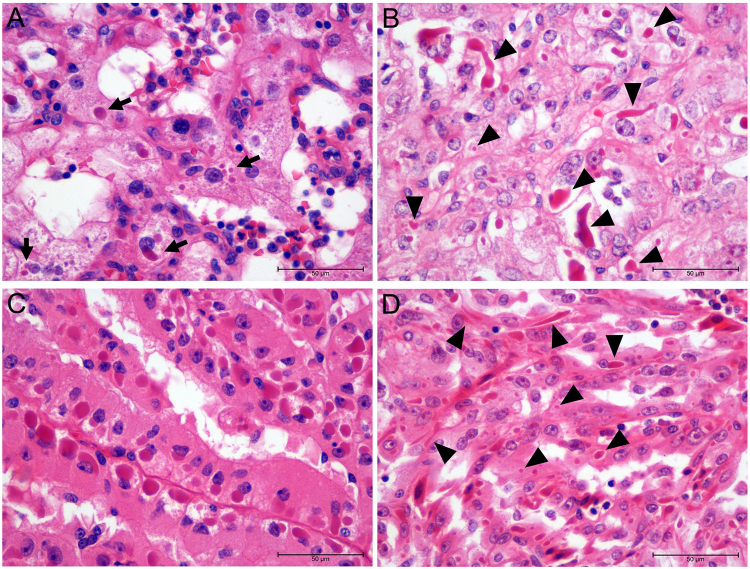
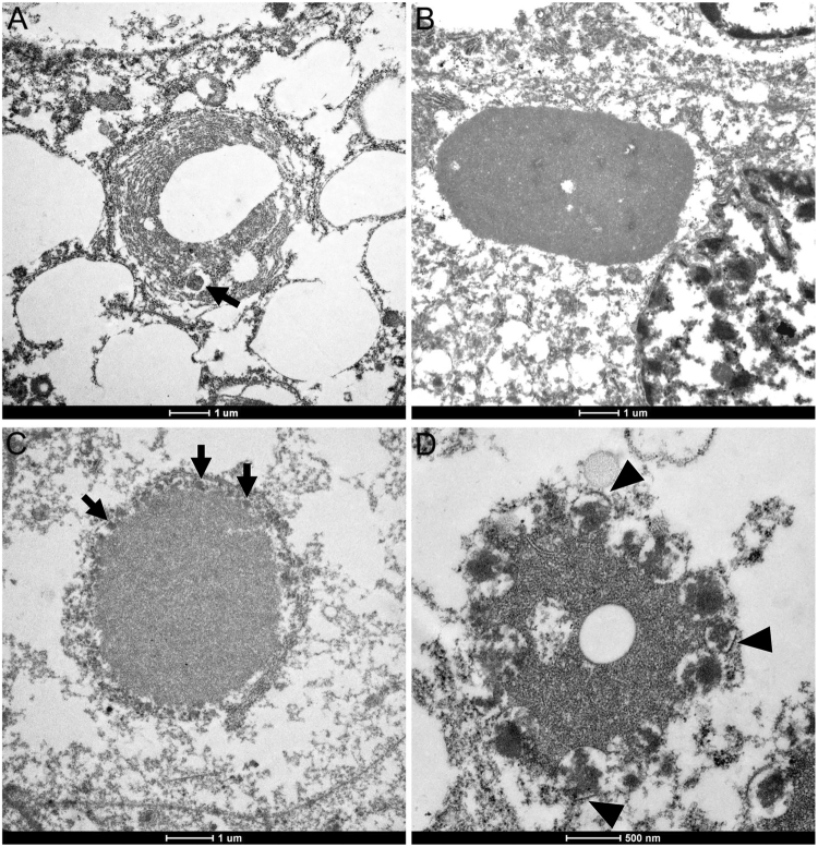
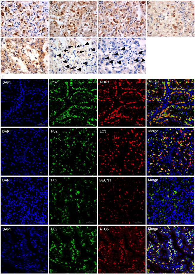
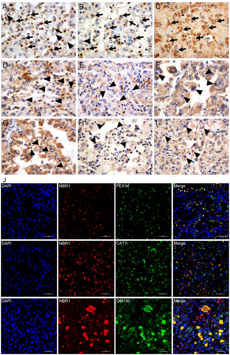
The relationship between autophagy and tumour is well studied, but tumour cell morphological changes associated with autophagy defects are rarely reported, especially in renal cell carcinoma (RCC). We collected 10 renal tumour samples with characteristic eosinophilic cytoplasmic inclusions (ECIs) and found that the ECIs were majorly composed of sequestosome 1/P62, neighbor of BRCA1 gene 1 (NBR1), PEX14, and CATALASE1 (CAT1). Further, transmission electron microscopy analysis revealed that ECIs were aggregates of proteinaceous material and peroxisomes. These results confirmed that ECIs in RCCs were the products of autophagy defects. The presence of ECIs was correlated with high Fuhrman grade components of RCCs. Whole-exome sequencing (WES) and Sanger sequencing confirmed that tumours with ECIs showed somatic mutations or high frequency of genetic variations in autophagy-related (ATG) genes, such as ATG7, ATG5, and ATG10. These results indicate that nucleotide changes in ATG genes are associated with autophagy defect, ECI formation, and even tumour grade in RCCs.


Conflict of interest statement

The authors declare no competing interests.

Figures

Figure 1
Figure 1
Light microscopic characteristics of ECIs in three types of RCCs (H&E, ×400). (A and B) ECIs in ccRCC. Note the variations in the number of ECIs (arrows) and their size (arrowheads) and shape (arrowheads). (C) Round-to-oval ECIs were observed to be regularly arranged in a juxtanuclear position in PRCC cells. (D) ECIs of varying sizes and shapes (arrowheads) were found in MTSCC cells.
Figure 2
Figure 2
Transmission electron micrographs of ECIs in RCCs. (A) An ECI exhibiting a concentric membranous structure with lipid or clear spaces; note the round electron-dense structure within the concentric membranous structure (arrow). (B) An ECI consisting of proteinaceous material with several clear spaces within it. (C) An ECI comprising proteinaceous material with electron-dense structures (arrows) completely enclosing it. (D) An ECI displaying aggregates of fibrillar components and single membrane-bound electron-dense structures (arrowheads).
Figure 3
Figure 3
Immunohistochemical staining and double-immunofluorescence labeled microscopic images of ECIs in RCCs. Immunohistochemical staining showed that the ECIs were immunopositive for P62 (A), NBR1 (B), LC3 (C), BECN1 (D), and ATG5 (E), and immunonegative for LCK (arrows in G). Ub1 immunopositivity was detected in a small number of ECIs (arrows in F). Double-immunofluorescence staining of ECIs demonstrated co-localization of P62 with NBR1, LC3, BECN1 or ATG5 in ECIs (H).
Figure 4
Figure 4
Localization of organelle markers in ECIs of RCCs. Immunohistochemical staining showed that ECIs were intensively positive for PEX14 and CAT1 (two markers of peroxisomes, arrows in A and B). Some of the ECIs were positive for GM130 (a marker of Golgi apparatus, arrows in C). None of the ECIs exhibited immunoreactivity of LAMP1 and LAMP2 (two markers of lysosome or autolysosome, arrowheads in D and E), TOM20 (a marker of mitochondria, arrowheads in F), CALR (a marker of the endoplasmic reticulum, arrowheads in G), RPS6 (a marker of ribosomes, arrowheads in H), or RAB5A (a marker of early endosomes, arrowheads in I). Double- immunofluorescence labelling of ECIs revealed that most of the NBR1-positive ECIs were positive for PEX14 and CAT1, and that a portion of them displayed moderate GM130 staining (J).
Figure 5
Figure 5
Three-dimensional reconstruction images of ECIs. (A) ECIs without NBR1 and peroxisomes. (AD) ECIs comprising P62, BECN1, and NBR1 with peroxisomes partially or completely surrounding them.

References

    1. Rini BI, Campbell SC, Escudier B. Renal cell carcinoma. Lancet. 2009;373:1119–1132. doi: 10.1016/S0140-6736(09)60229-4. - DOI - PubMed
    1. Jagirdar J, et al. Globular Mallory-like bodies in renal cell carcinoma: report of a case and review of cytoplasmic eosinophilic globules. Hum Pathol. 1985;16:949–952. doi: 10.1016/S0046-8177(85)80135-0. - DOI - PubMed
    1. Unger P, et al. Eosinophilic globules resembling Mallory bodies in a renal cell carcinoma. N Y State J Med. 1992;92:18–20. - PubMed
    1. Gatalica Z, Miettinen M, Kovatich A, McCue PA. Hyaline globules in renal cell carcinomas and oncocytomas. Hum Pathol. 1997;28:400–403. doi: 10.1016/S0046-8177(97)90026-5. - DOI - PubMed
    1. Krishnan B, Truong LD. Renal epithelial neoplasms: the diagnostic implications of electron microscopic study in 55 cases. Hum Pathol. 2002;33:68–79. doi: 10.1053/hupa.2002.30210. - DOI - PubMed

Publication types

MeSH terms