. 2018 Aug;24(8):1121-1127.
doi: 10.1038/s41591-018-0087-6. Epub 2018 Jul 2.

Metformin reverses established lung fibrosis in a bleomycin model

Affiliations

Metformin reverses established lung fibrosis in a bleomycin model

Sunad Rangarajan et al. Nat Med. 2018 Aug.

Erratum in

Abstract

Fibrosis is a pathological result of a dysfunctional repair response to tissue injury and occurs in a number of organs, including the lungs1. Cellular metabolism regulates tissue repair and remodelling responses to injury2-4. AMPK is a critical sensor of cellular bioenergetics and controls the switch from anabolic to catabolic metabolism5. However, the role of AMPK in fibrosis is not well understood. Here, we demonstrate that in humans with idiopathic pulmonary fibrosis (IPF) and in an experimental mouse model of lung fibrosis, AMPK activity is lower in fibrotic regions associated with metabolically active and apoptosis-resistant myofibroblasts. Pharmacological activation of AMPK in myofibroblasts from lungs of humans with IPF display lower fibrotic activity, along with enhanced mitochondrial biogenesis and normalization of sensitivity to apoptosis. In a bleomycin model of lung fibrosis in mice, metformin therapeutically accelerates the resolution of well-established fibrosis in an AMPK-dependent manner. These studies implicate deficient AMPK activation in non-resolving, pathologic fibrotic processes, and support a role for metformin (or other AMPK activators) to reverse established fibrosis by facilitating deactivation and apoptosis of myofibroblasts.


Conflict of interest statement

Disclosures

All authors declare no conflict of interest.

Figures

Figure 1
Figure 1
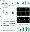
Distinct patterns of AMPK activity in lung epithelial cells and myofibroblasts of human individuals with IPF. (a) Representative images show pT172-AMPK (red), epithelial marker T1α (green), α-SMA (green) and nuclei (blue) in lung sections of control and subjects with IPF. Scale bar, 100 µm. Right panels display magnified areas from images indicated by dashed boxes. Scale bar, 30 µm. (b) Scattergrams indicate fluorescence intensity and Pearson’s correlation (r) in images display in a. (c) The ratio of pT172-AMPK, T1α or α-SMA to nuclei fluorescence intensity. Means ± SD; n = 9 (control), n = 9 (IPF) for both pT172-AMPK and α-SMA; n = 8 (control), n = 6 (IPF) for T1α. *P < 0.05 (Student’s t-test). (d) Representative western blots and quantitative analysis show the amounts of pT172-AMPK, pS240/244-S6 kinase, collagen type I, fibronectin, LC3BI/II and β-actin in IPF fibroblasts stimulated with AICAR (24 h). LC3II/I ratio was obtained from cells treated with AICAR and chloroquine. Means ± SD, n = 3 or n = 4. *P < 0.05 (ANOVA).
Figure 2
Figure 2
AMPK activation reduces the levels of ECM proteins in TGF-β1-treated fibroblasts. (a) Representative western blots show collagen, fibronectin, pT172-AMPK, pS423/425-SMAD3 and β-actin in human lung fibroblasts treated with TGF-β1 for 24 hours, followed by AICAR for an additional 24 hours. Means ± SD, n = 3. *P < 0.05 (ANOVA). (b) Levels of collagen decrease in fibroblasts treated with metformin- (0 or 1 mM; 24 hours) in TGF-β1-differrentiated myofibroblasts. Representative immunoblots are shown. Means ± SD, n = 3. *P < 0.05 (ANOVA). (c,d) Representative images of collagen and LC3 indicate fluorescent staining in control and chloroquine-treated MEFs. Collagen (green), LC3 (red), nuclei (blue). Scale bar, 25 µM. (d) High magnification areas are selected by dashed boxes depicted in c. Scale bar, 10 µM. (e) Representative western blots show the levels of collagen accumulation in control (scrambled siRNA) and human lung fibroblasts with siRNA-mediated silencing of beclin, LC3B or AMPK. Cells were treated TGF-β1 for 24 hours and then AICAR for an additional 24 hours. (f) Optical band densitometry of immunoblots in e. Means ± SD, n = 3. *P < 0.05, ns-not significant (ANOVA). #P < 0.05 (Student’s t-test).
Figure 3
Figure 3
Effects of AMPK activation on mitochondrial bioenergetics and TGF-β1-mediated resistance to apoptosis in lung fibroblasts. (a) Representative transmission electron micrographs show mitochondrial size in cross-section of fibroblasts from control and subjects with IPF. Arrows (mitochondria), CR (cristae), C (cytoplasm), G (golgi). Scale bar, 200 nm. Means ± SEM, n = 21 (control), n = 37 (IPF) of mitochondria morphometric analysis. *P < 0.05 (Student’s t-test). (b) Representative OCR tracing (left) and bioenergetic indices (right) from wild-type and AMPKα1/2−/− MEFs. Means ± SD, n = 5. *P < 0.05 (Student’s t-test). (c) Representative western blots of 555-ULK1 phosphorylation in wild-type and AMPK-deficient MEFs. (d) Representative fluorescent staining images of TUNEL (green) and nuclei (blue) in human lung fibroblasts treated with TGF-β1 for 24 hours, followed by AICAR for an additional 7 days. Red arrows indicate TUNEL positive cells. Scale bar, 50 µm. (e) Representative immunoblots of cleaved PARP, Bcl2, p53, and β-actin from fibroblasts treated as indicated in d. (f) Representative immunoblots and quantitative analysis of cleaved PARP in human lung fibroblasts treated with TGF-β1 for 24 hours, followed by AICAR (72 hours), and then antimycin A (AA; 16 hours). Means ± SD, n = 3. *P < 0.05 (ANOVA). (g) Representative immunoblots of TFAM and components of the ETC complexes in human lung fibroblasts treated with AICAR or metformin for 72 hours. Means ± SD, n = 3. *P < 0.05 (ANOVA).
Figure 4
Figure 4
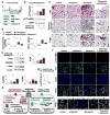
Metformin accelerates resolution of bleomycin-induced lung fibrosis. Panel (a) outlines the design of therapeutic dosing of metformin in mice with established fibrosis following bleomycin-induced lung injury. (b) Representative western blots indicate the amounts of pT172-AMPK and total AMPK in lung homogenates from mice treated as depicted in a. Means ± SD, n = 3. *P < 0.05 (ANOVA). (c) Representative images show H&E, Masson’s trichrome and α-SMA staining of lung sections from indicated groups of mice. Scale bar, 100 µm. (d) Quantitative analysis of hydroxyproline and TGF-β1 in lung homogenates from groups of mice depicted in a. Means ± SD, n = 5. *P < 0.05 (ANOVA). (e) Representative immunoblots of TFAM, NDUFB8 and α-SMA in whole lung homogenates of mice as indicated. Means ± SD, n = 3. *P < 0.05 (ANOVA). (f) Representative fluorescence images of pT172-AMPK and T1α in lung sections of mice treated, as depicted in a. pT172-AMPK (red), type I alveolar epithelial cell marker T1α (green), nuclei (blue). Scale bar, 50 µm. Lower panel shows higher magnification images of areas marked by square boxes (dash lines). Scale bar, 10 µm. (g) Quantitative analysis of pT172-AMPK/nuclei fluorescence ratio from normal and fibrotic areas of bleomycin-injured mice. Means ± SD, 3 mice per group, n = 9 normal areas, n = 9 active fibrosis. *P < 0.05 (Student’s t-test). (h) Schematic diagram of the proposed mechanisms for fibrosis resolution induced by AMPK activation. Left panel: loss of AMPK activity promotes persistent myofibroblast activation by deficient autophagy, ECM accumulation, mitochondrial dysfunction and acquired apoptosis resistance. Right panel: AMPK activation stimulates autophagy and facilitates ECM turnover, while inducing mitochondrial biogenesis, thus restoring sensitivity to apoptosis and promoting fibrosis resolution.

Comment in

References

    1. Wynn TA, Ramalingam TR. Mechanisms of fibrosis: therapeutic translation for fibrotic disease. Nat Med. 2012;18:1028–1040. - PMC - PubMed
    1. Lumeng CN, Saltiel AR. Inflammatory links between obesity and metabolic disease. J Clin Invest. 2011;121:2111–2117. - PMC - PubMed
    1. O'Neill LA, Hardie DG. Metabolism of inflammation limited by AMPK and pseudo-starvation. Nature. 2013;493:346–355. - PubMed
    1. Eltzschig HK, Eckle T. Ischemia and reperfusion--from mechanism to translation. Nat Med. 2011;17:1391–1401. - PMC - PubMed
    1. Hardie DG, Ross FA, Hawley SA. AMPK: a nutrient and energy sensor that maintains energy homeostasis. Nat Rev Mol Cell Biol. 2012;13:251–262. - PMC - PubMed

Publication types