. 2018 Jul 2;8(1):9975.
doi: 10.1038/s41598-018-28195-2.

Somatosensory innervation of the oral mucosa of adult and aging mice

Affiliations

Somatosensory innervation of the oral mucosa of adult and aging mice

Yalda Moayedi et al. Sci Rep. .

Abstract

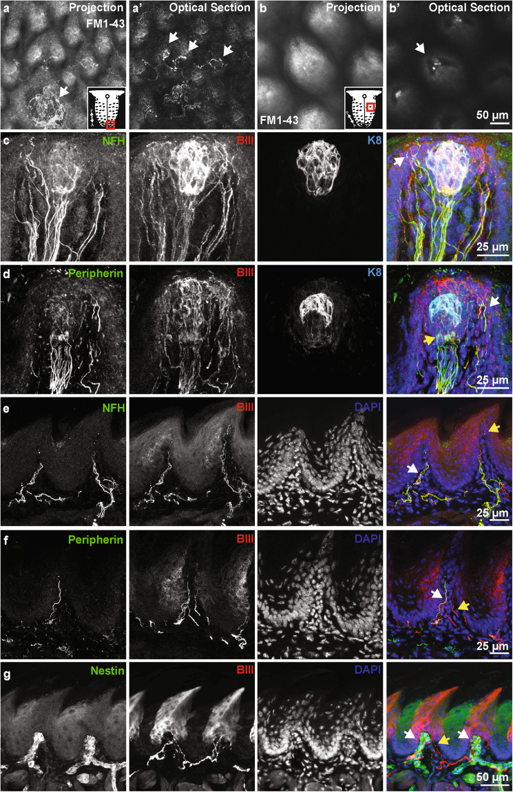
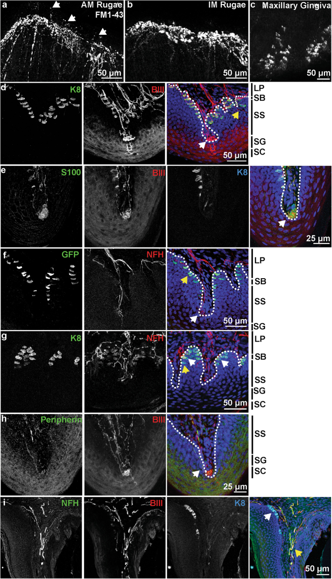
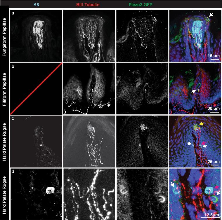
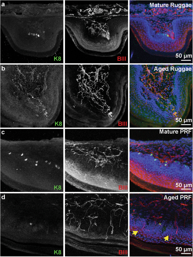
Oral mechanoreception is implicated in fundamental functions including speech, food intake and swallowing; yet, the neuroanatomical substrates that encode mechanical stimuli are not well understood. Tactile perception is initiated by intricate mechanosensitive machinery involving dedicated cells and neurons. This signal transduction setup is coupled with the topology and mechanical properties of surrounding epithelium, thereby providing a sensitive and accurate system to detect stress fluctuations from the external environment. We mapped the distribution of anatomically distinct neuronal endings in mouse oral cavity using transgenic reporters, molecular markers and quantitative histomorphometry. We found that the tongue is equipped with an array of putative mechanoreceptors that express the principal mechanosensory channel Piezo2, including end bulbs of Krause innervating individual filiform papillae and a novel class of neuronal fibers innervating the epithelium surrounding taste buds. The hard palate and gums are densely populated with three classes of sensory afferents organized in discrete patterns including Merkel cell-neurite complexes, Meissner's corpuscles and glomerular corpuscles. In aged mice, we find that palatal Merkel cells reduce in number at key time-points that correlate with impaired oral abilities, such as swallowing and mastication. Collectively, this work identifies the mechanosensory architecture of oral tissues involved in feeding.


Conflict of interest statement

Portions of this work were funded by NesTec.

Figures

Figure 1
Figure 1
The lingual mucosa is innervated by multiple neuronal classes. (a) Whole mount imaging of FM1-43 labeling of neuronal afferents in the tongue. A projection shows neuronal endings surrounding a fungiform papillae in the anterior tongue. An optical section through this image reveals neuronal endings innervating individual filiform papillae (a’, arrows). Schematic shows tongue region represented. (b) Whole mount imaging of filiform papillae in posterior tongue. An optical section through the base of the papillae (b’) reveals neuronal afferents innervating the core of individual papillae (arrow). Schematic shows tongue region represented. (c) Neurofilament heavy (NFH) positive and negative fibers innervate fungiform papillae. NFH fibers extend into the epidermis overlying the taste cells (arrow). (d) Peripherin+ fibers also enter the fungiform papillae, these primarily associate with taste cells (yellow arrow, K8), but also extend around to the overlying epithelium (white arrow). (e) Filiform papillae are innervated with NFH+ afferents (white arrow). Note other afferents extend into the apical regions of the papilla (yellow arrow). (f) Peripherin+ (white arrow) and Peripherin- (yellow arrow) afferents are also present in the filiform papilla, demonstrating that multiple neuronal subtypes innervate these non-taste papillae. (g) A subset of neuronal endings in the filiform papillae are surrounded by Nestin+ cells.
Figure 2
Figure 2
Maxillary epithelia are rich in neuronal innervation and Merkel cells. (a) Whole mount imaging of FM1-43 labeling in an antemolar ruga of the hard palate. Dense innervation lines the ridge of the antemolar ruga, with ultraterminals jutting into the epithelium of the ruga (arrows). (b) FM1-43 labels dense neuronal endings in the ridges of intermolar ruga. (c) FM1-43 labels Merkel cell clusters in maxillary gingiva. (d)Innervation of hard palate rugae was visulaized with βIII tubulin and K8. Glomerular-type endings were embedded in epithelial pegs (white arrow), often complemented with an ultra-terminal (asterisk). Free nerve fibers innervated the epithelium (yellow arrow). K8 staining showed Merkel cells densely packed along the epithelial-lamina propria junction. (e) Glomerular endings (arrow) associated with end-terminal Schwann cells, visualized by S100 staining. (f) NFH revealed neuronal endings with Meissner’s morphology (white arrow). This was also associated with an ultra-terminal (asterisk). Atoh1-GFP+ Merkel cells were found lining the epithelial ridges. Yellow arrow denotes a Merkel cell that is not innervated by a NFH+ afferent. (g) Coronal section of a hard palate ridge shows K8+ Merkel cells organized in epithelial pegs. Merkel cells innervated by NFH+ afferents (white arrow) as well as Merkel cells without NFH+ neuronal connections (yellow arrow) are denoted. (h) Peripherin+ endings were found innervating palatal ridges (arrow). These did not appear to be a large component of the glomerular-type endings. Merkel cells line exterior aspect of maxillary gingiva mucosa (white arrow), visualized by coronal sections and immunohistochemistry. NFH+ neurons innervate the gingiva as well (yellow arrow); however, these did not appear to form organized end organs. (LP: lamina propria, SB: stratum basale, SS: stratum spinosum, SG: stratum granulosum, SC: stratum corneum, Dotted line marks LP-SB border).
Figure 3
Figure 3
Piezo2 localizes to subsets of neuronal endings in the tongue and hard palate. (a) Piezo2 protein was found in sensory endings that surround taste cells in fungiform papillae (arrow). These endings form bulbous end feet that innervate the epidermis. (b) A subset of neuronal endings in filiform papillae were also Piezo2+. These likely represent end bulbs of Krause (arrow). (c) In the hard palate rugae, Piezo2 was localized to glomerular endings (yellow arrow), as well as in K8+ Merkel cells (red arrow). Piezo2+ crescent-shaped structures in the same region could represent K8- Merkel cells or adjacent sensory neuronal terminals (white arrow). (d) Expanded image of a Piezo2+, K8+ Merkel cell and Piezo2+, K8-.
Figure 4
Figure 4
Merkel cell density reduces with age in maxillary epithelia. (a) Atoh1LacZ/+ hard palate reveals a high density of Merkel cells lining rugae. Merkel cells were particularly dense in the incisive papilla, lining the teeth, and in the postrugal field. (b) Aged hard palates reveal a drastic reduction in the density of Merkel cells. Note that distinct pinprick punctae are replaced with blobs of LacZ staining in some regions. (c) Merkel cells were found to line the maxillary gums in a discrete stripe of cells in mature mice. (d) In aged mice, the distinct line of Merkel cells is abolished. (e) Quantification of LacZ density in the hard palate reveals reductions in Merkel cells with aging (N = 3–4 mice). Schematics reveal decreases in the density of LacZ+ staining in antemolar and intermolar rugae. Two-way ANOVA reveals significant differences between mature (7–15 weeks) and aged (12.5–20 months) palates ( p = 0.0009) and between rugae and interrugal epithelium (IRE) ( p < 0.0001). Bonferroni posthoc analysis found significant reductions in the density of LacZ staining with age in rugal epithelium but not between IRE. ** p< 0.01 H&E staining of mature palate reveals structure of rugae. Blue regions are areas of LacZ+ staining. Note, stratum corneum separated during mounting process, a routine occurance in sectioning cornified tissue. (f) High magnification of a mature rugae shows epithelial pegs and areas of LacZ-stained epithelia. (g) Aged palate structure remains intact, with prominent rugae. Note reduction in blue LacZ+ regions compared with f. (h) High magnification of aged rugae reveals intact epithelium.
Figure 5
Figure 5
Innervation of the tongue and hard palate with age. (a) The posterior rugae from a mature mouse displays dense Merkel cells. (b) Aged posterior rugae displays no Merkel cells. (c) The postrugal field from a mature mouse has abundant Merkel cells, frequent neuronal endings in the lamina propria and few epidermal fibers. (d) The postrugal field from an aged mouse has infrequent Merkel cells, less dense neuronal innervation in the lamina propria and frequent epidermal fibers (yellow arrows).
Figure 6
Figure 6
Summary of oral mechanoreceptor anatomy. Schematic of presumptive somatosensory innervation of the lingual and palatal mucosa. Myelinated afferents are shown in blue and marked by βIII tubulin and NFH; these endings are generally FM1-43+ and expected to be Piezo2+. Unmyelinated afferents are shown in pink and marked by expression of βIII tubulin and Peripherin, these are largely FM1-43- and Piezo2-. The lingual mucosa is innervated by two presumptive mechanosensory end organs. End bulbs of Krause are found in filiform papillae, while a neuronal collar surrounds taste buds in filiform papillae. In addition to these, unmyelinated afferents extend into the filiform papillae and surround taste buds. The hard palate is innervated by glomerular endings and corpuscular endings. These end organs are morphologically distinguished by the relatively disorganized appearance of glomerular endings. The hard palate rugae and post-rugal field are also densely lined with Merkel cell-neurite complexes. Finally, unmyelinated afferents are interspersed throughout the palatal epithelium. Cartoon was drawn with color schemes and symbols modeled after Fig. 1 of Bautista & Lumpkin.

Similar articles

Cited by

References

    1. Mouritsen OG. Gastrophysics of the Oral Cavity. Curr Pharm Des. 2016;22:2195–2203. doi: 10.2174/1381612822666160219120940. - DOI - PubMed
    1. Mars M, Hogenkamp PS, Gosses AM, Stafleu A, De Graaf C. Effect of viscosity on learned satiation. Physiol Behav. 2009;98:60–66. doi: 10.1016/j.physbeh.2009.04.018. - DOI - PubMed
    1. Dominy NJ, et al. How chimpanzees integrate sensory information to select figs. Interface Focus. 2016;6:20160001. doi: 10.1098/rsfs.2016.0001. - DOI - PMC - PubMed
    1. Zijlstra N, Mars M, de Wijk RA, Westerterp-Plantenga MS, de Graaf C. The effect of viscosity on ad libitum food intake. Int J Obes (Lond) 2008;32:676–683. doi: 10.1038/sj.ijo.0803776. - DOI - PubMed
    1. Zijlstra N, et al. Effect of viscosity on appetite and gastro-intestinal hormones. Physiol Behav. 2009;97:68–75. doi: 10.1016/j.physbeh.2009.02.001. - DOI - PubMed

Publication types

MeSH terms