. 2018 Jul 2;8(1):9914.
doi: 10.1038/s41598-018-28220-4.

Supraphysiological Levels of Oxygen Exposure During the Neonatal Period Impairs Signaling Pathways Required for Learning and Memory

Affiliations

Supraphysiological Levels of Oxygen Exposure During the Neonatal Period Impairs Signaling Pathways Required for Learning and Memory

Manimaran Ramani et al. Sci Rep. .

Abstract

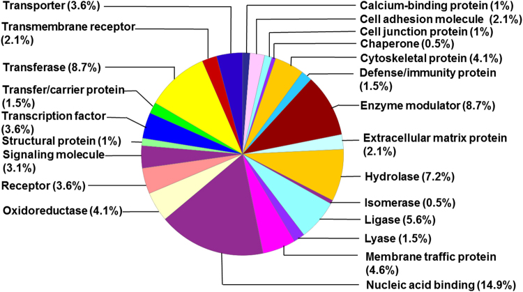
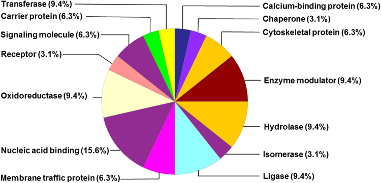
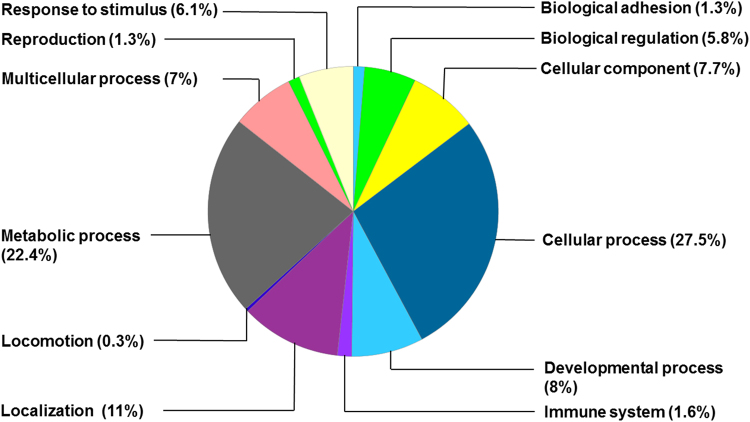
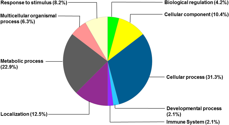
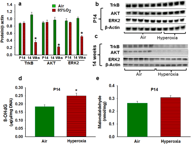
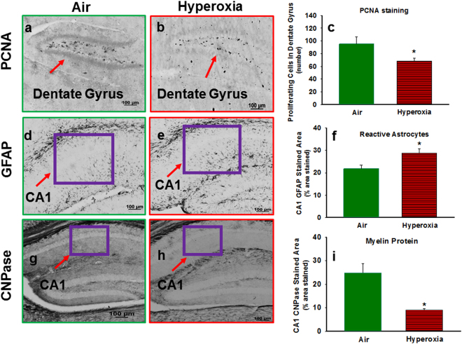
Preterm infants often require prolonged oxygen supplementation and are at high risk of neurodevelopmental impairment. We recently reported that adult mice exposed to neonatal hyperoxia (postnatal day [P] 2 to 14) had spatial navigation memory deficits associated with hippocampal shrinkage. The mechanisms by which early oxidative stress impair neurodevelopment are not known. Our objective was to identify early hyperoxia-induced alterations in hippocampal receptors and signaling pathways necessary for memory formation. We evaluated C57BL/6 mouse pups at P14, exposed to either 85% oxygen or air from P2 to 14. We performed targeted analysis of hippocampal ligand-gated ion channels and proteins necessary for memory formation, and global bioinformatic analysis of differentially expressed hippocampal genes and proteins. Hyperoxia decreased hippocampal mGLU7, TrkB, AKT, ERK2, mTORC1, RPS6, and EIF4E and increased α3, α5, and ɤ2 subunits of GABAA receptor and PTEN proteins, although changes in gene expression were not always concordant. Bioinformatic analysis indicated dysfunction in mitochondria and global protein synthesis and translational processes. In conclusion, supraphysiological oxygen exposure reduced proteins necessary for hippocampus-dependent memory formation and may adversely impact hippocampal mitochondrial function and global protein synthesis. These early hippocampal changes may account for memory deficits seen in preterm survivors following prolonged oxygen supplementation.


Conflict of interest statement

The authors declare no competing interests.

Figures

Figure 1
Figure 1
Distribution of upregulated hippocampal proteins by class in the hyperoxia-exposed group.
Figure 2
Figure 2
Distribution of downregulated hippocampal proteins by class in the hyperoxia-exposed group.
Figure 3
Figure 3
Distribution of upregulated hippocampal proteins by biological processes in the hyperoxia-exposed group.
Figure 4
Figure 4
Distribution of downregulated hippocampal proteins by biological processes in the hyperoxia-exposed group.
Figure 5
Figure 5
Effect hyperoxia on whole hippocampal TrkB, AKT, and ERK2 concentrations and oxidative stress markers. (a) Graphical representative of Western blot results from hippocampal samples of P14 and 14 weeks old mice. At P14, total TrkB, AKT, and ERK2 levels were not significantly different between air-and hyperoxia-exposed mice. At 14 weeks, TrkB, AKT, and ERK2 levels were decreased in the hyperoxia group. (b) Representative of hippocampal western blots from P14 mice exposed to Air (n = 6) or Hyperoxia (n = 4). The blots were obtained from different gels and pictures were taken with the same exposure. (c) Representative of hippocampal western blots from 14-week-old mice exposed to Air (n=) or Hyperoxia (n = 6). The blots were obtained from different gels and pictures were taken with the same exposure. (d) Graphical representative of 8-OHdG (oxidative DNA damage marker) ELISA results from P14 mice exposed to Air (n=) or Hyperoxia (n = 6). Hyperoxia-exposed mice had increased 8-OHdG in the hippocampus. (e) Graphical representative of malodialdehyde (MDA adducts: lipid peroxidation marker) ELISA results from P14 mice exposed to Air (n = ) or Hyperoxia (n = 6). No difference in MDA adducts were observed between the groups. In the panels a, d, and e; Air: solid green bars, Hyperoxia: solid red bars with horizontal stripes. *Represents p < 0.05; Air vs. Hyperoxia.
Figure 6
Figure 6
Histological assessment of effect hyperoxia on cell proliferation, reactive astrocytes and myelin protein. Photomicrographs of PCNA stained brain sections from P14 mice exposed to Air (a) or Hyperoxia (b). (c) Graphical representation of PCNA staining results; hyperoxia-exposed mice had a reduction in the number of proliferative cells in the dentate gyrus. Photomicrographs of GFAP stained brain sections from P14 mice exposed to Air (d) or Hyperoxia (e). (f) Graphical of representation GFAP staining results; hyperoxia-exposed mice had an increased in the percentage of GFAP stained area in the CA1 region. Photomicrographs of CNPase stained brain sections from P14 mice exposed to Air (g) or Hyperoxia (h). (i) Graphical representation of CNAPase staining results; hyperoxia exposed mice had decreased in the percentage of CNPase stained area in the CA1 region of the hippocampus. In the panels (c,f), and (i); Air: solid green bars, Hyperoxia: solid red bars with horizontal stripes. *Represents p < 0.05; Air vs. Hyperoxia.

Similar articles

Cited by

References

    1. Aarnoudse-Moens CS, Weisglas-Kuperus N, van Goudoever JB, Oosterlaan J. Meta-analysis of neurobehavioral outcomes in very preterm and/or very low birth weight children. Pediatrics. 2009;124:717–728. doi: 10.1542/peds.2008-2816. - DOI - PubMed
    1. Luu TM, Ment L, Allan W, Schneider K, Vohr BR. Executive and memory function in adolescents born very preterm. Pediatrics. 2011;127:e639–646. doi: 10.1542/peds.2010-1421. - DOI - PMC - PubMed
    1. Omizzolo C, et al. Neonatal brain abnormalities and memory and learning outcomes at 7 years in children born very preterm. Memory. 2014;22:605–615. doi: 10.1080/09658211.2013.809765. - DOI - PMC - PubMed
    1. Gimenez M, et al. Hippocampal gray matter reduction associates with memory deficits in adolescents with history of prematurity. NeuroImage. 2004;23:869–877. doi: 10.1016/j.neuroimage.2004.07.029. - DOI - PubMed
    1. Nicolaides KH, Soothill PW, Rodeck CH, Campbell S. Ultrasound-guided sampling of umbilical cord and placental blood to assess fetal wellbeing. Lancet. 1986;1:1065–1067. doi: 10.1016/S0140-6736(86)91333-4. - DOI - PubMed

Publication types