. 2018 Jul;25(7):616-622.
doi: 10.1038/s41594-018-0085-x. Epub 2018 Jul 2.

Structure of the Cdc48 ATPase with its ubiquitin-binding cofactor Ufd1-Npl4

Affiliations

Structure of the Cdc48 ATPase with its ubiquitin-binding cofactor Ufd1-Npl4

Nicholas O Bodnar et al. Nat Struct Mol Biol. 2018 Jul.

Abstract

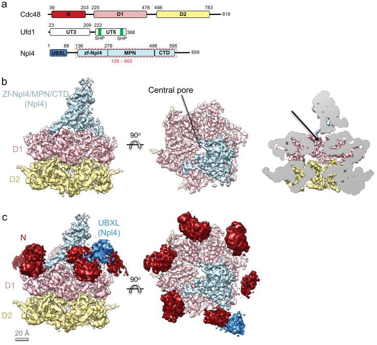
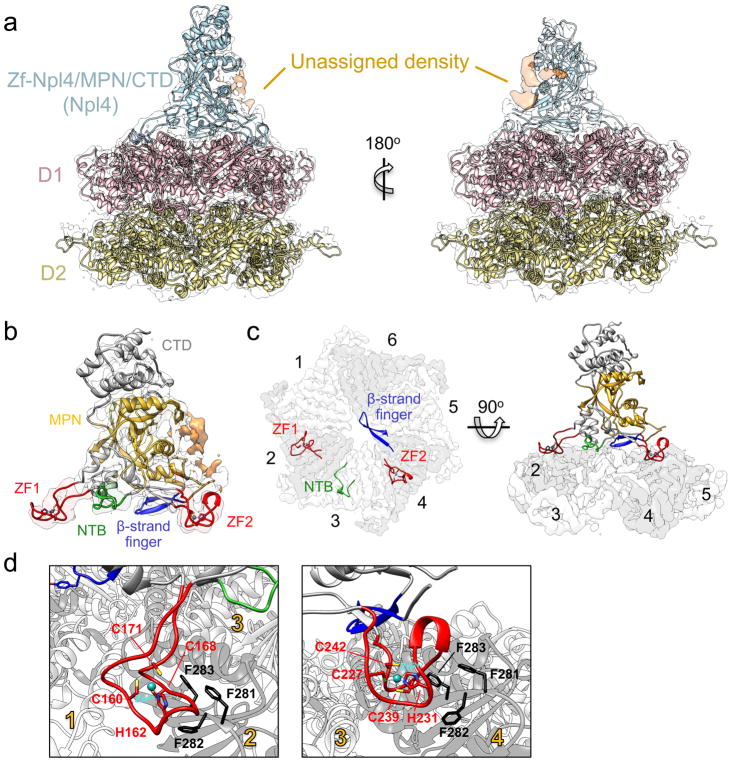
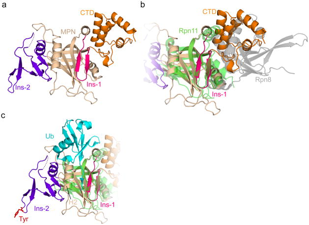
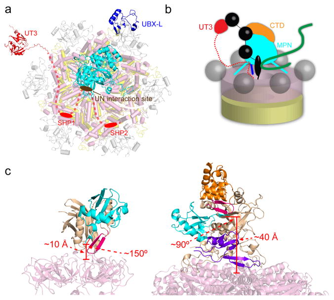
Many polyubiquitinated proteins are extracted from membranes or complexes by the conserved ATPase Cdc48 (in yeast; p97 or VCP in mammals) before proteasomal degradation. Each Cdc48 hexamer contains two stacked ATPase rings (D1 and D2) and six N-terminal (N) domains. Cdc48 binds various cofactors, including the Ufd1-Npl4 heterodimer. Here, we report structures of the Cdc48-Ufd1-Npl4 complex from Chaetomium thermophilum. Npl4 interacts through its UBX-like domain with a Cdc48 N domain, and it uses two Zn2+-finger domains to anchor the enzymatically inactive Mpr1-Pad1 N-terminal (MPN) domain, homologous to domains found in several isopeptidases, to the top of the D1 ATPase ring. The MPN domain of Npl4 is located above Cdc48's central pore, a position similar to the MPN domain from deubiquitinase Rpn11 in the proteasome. Our results indicate that Npl4 is unique among Cdc48 cofactors and suggest a mechanism for binding and translocation of polyubiquitinated substrates into the ATPase.


Conflict of interest statement

The authors declare no competing financial interests.

Figures

Figure 1
Figure 1. Structure of the Cdc48/UN complex with ATPγS
a, Domain organization of Chaetomium thermophilum Cdc48 and its co-factors, Ufd1 and Npl4. The Npl4 region indicated with a red dashed box was crystallized. b, Cryo-EM density map of the Cdc48/UN complex, colored as in a. The N domains were masked out in the final refinement step. The rightmost panel shows a side cutaway view. The arrow indicates the putative path of the substrate into the pore. c, The N domains of Cdc48 (dark red) from a map refined without a mask are shown relative to the map obtained with masking. The contiguous extra density next to one of the N domains (dark blue) is assigned to the UBX-like domain of Npl4.
Figure 2
Figure 2. Interactions between Cdc48 and its cofactor
a, A crystal structure of Npl4 (light blue) and a homology model of Cdc48 (D1 in pink; D2 in yellow) were docked into the cryo-EM map. Unassigned density (orange) likely belongs to parts of the UT6 domain of Ufd1. b, Close-up view of the Npl4 crystal structure docked into the cryo-EM map. The MPN domain is shown in yellow and D1-interacting regions extending from it are highlighted: the two Zn2+ fingers (ZF; red), an N-terminal bundle (NTB; light green), and the ‘β-strand finger’ (blue). c, Top view of D1-interacting regions, colored as in b, with the D1 ring shown as a white/gray surface. For clarity, D2 was omitted. ATPase subunits of the Cdc48 hexamer are numbered. d, Close-up views of the Zn2+ fingers, with Zn2+-coordinating residues in stick representation. The interacting tri-phenylalanine (FFF) sequence in Cdc48 is highlighted.
Figure 3
Figure 3. Functional analysis of Zn2+-finger and MPN domain mutations
a, Unfolding of poly-ubiquitinated Eos by wild-type S. cerevisiae Cdc48 and the indicated UN variants. ZF1: H139A/C145A. ZF2: H208A/C216A. ZF1/2: H139A/C145A/H208A/C216A. Data are shown as mean ± SD of n=3 technical replicates. See Suppl Data Set 1. b, A npl4-1 temperature-sensitive S. cerevisiae strain was transformed with a plasmid encoding wild-type Npl4 or the indicated Zn2+-finger mutants, spotted in serial dilution, and incubated at the indicated temperatures for two days (30 and 37°C) or three days (25°C). c, As in a, but with mutants in the FFF motif (residues 275-277) of Cdc48. Data are shown as mean ± SD of n=3 technical replicates. See Suppl Data Set 1. d, Binding of poly-ubiquitinated substrate to SBP-tagged C. thermophilum Npl4 (Zn2+-finger/MPN/CTD domains, residues 129-602) or the indicated variants (MPN only: residues 129-519, CTD only: residues 519-602). The bait proteins were bound to streptavidin beads and incubated with dye-labeled, poly-ubiquitinated superfolder GFP. Bound material was analyzed by SDS-PAGE followed by fluorescence scanning (top) and Coomassie blue staining (bottom). M, molecular weight markers. e, The locations of MPN cleft mutants tested in d are shown in stick representation. The MPN, insert-1, insert-2, and CTD are shown in tan, magenta, purple, and orange, respectively.
Figure 4
Figure 4. MPN domain of Npl4
a, Crystal structure of the MPN/CTD domains of Npl4. The core MPN region is in tan, the insert-1 (Ins-1) region in magenta, the insert-2 (Ins-2) region in purple, and the CTD in orange. The dashed line indicates a 9-residue acidic loop unresolved in the crystal structure. b, As in a, but with the Rpn11/Rpn8 structure overlaid (PDB: 5U4P). Rpn11 is in green and Rpn8 in gray. c, As in a, with the Rpn11 MPN (green) and its associated ubiquitin (cyan) overlaid (PDB: 5U4P). The conserved Tyr at the tip of Ins-2 is shown in stick representation in red. H2: MPN helix 2.
Figure 5
Figure 5. Model for Cdc48/UN function
a, Arrangement of the various domains in the Cdc48/UN complex. The UBX-like (PDB: 2PJH) and Zn2+-finger/MPN domains of Npl4 are shown in dark blue and cyan, respectively. The UT3 domain of Ufd1 (in red; PDB: 1ZC1) is flexibly attached to the complex. The Npl4-interacting region of UT6 is shown as a brown oval, and the SHP boxes anchoring Ufd1 to the N domains of Cdc48 as red ovals. The intervening segments are shown as dashed lines. b, Model for the path of a poly-ubiquitinated substrate (black: ubiquitin; green: substrate protein) into the central pore of Cdc48. Four ubiquitin molecules are shown: one associated with UT3, one bound at the interface between CTD and MPN, one located with its C terminus in the MPN cleft, and one in the gap between MPN and the ATPase, which might serve as initiation site for translocation into the pore. c, Comparison between the location of the ubiquitin-bound MPN domain in the proteasome (left) with that predicted in the Cdc48 complex (right). For Rpn11, the core MPN region is in tan and the insert-1 (Ins-1) region in magenta. Ubiquitin is in cyan. For Npl4, the core MPN region is in tan, the insert-1 (Ins-1) region in magenta, the insert-2 (Ins-2) region in purple, and the CTD in orange. The approximate distance from the ubiquitin C terminus to the central pore opening and the approximate angle between the MPN cleft and the central pore axis are marked in red in each case. The proteasome/ubiquitin model was generated from PDBs 5T0H and 5U4P.

Similar articles

Cited by

References

    1. Stach L, Freemont PS. The AAA+ ATPase p97, a cellular multitool. Biochem J. 2017;474:2953–2976. - PMC - PubMed
    1. Zhang X, et al. Structure of the AAA ATPase p97. Mol Cell. 2000;6:1473–1484. - PubMed
    1. Meyer HH, Shorter JG, Seemann J, Pappin D, Warren G. A complex of mammalian ufd1 and npl4 links the AAA-ATPase, p97, to ubiquitin and nuclear transport pathways. EMBO J. 2000;19:2181–2192. - PMC - PubMed
    1. Lander GC, et al. Complete subunit architecture of the proteasome regulatory particle. Nature. 2012;482:186–191. - PMC - PubMed
    1. Tang WK, et al. A novel ATP-dependent conformation in p97 N-D1 fragment revealed by crystal structures of disease-related mutants. EMBO J. 2010;29:2217–2229. - PMC - PubMed

Publication types

MeSH terms

LinkOut - more resources