. 2018 Jul;25(7):583-590.
doi: 10.1038/s41594-018-0086-9. Epub 2018 Jul 2.

Structure of the core of the type III secretion system export apparatus

Affiliations

Structure of the core of the type III secretion system export apparatus

Lucas Kuhlen et al. Nat Struct Mol Biol. 2018 Jul.

Erratum in

Abstract

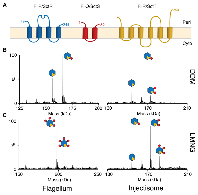
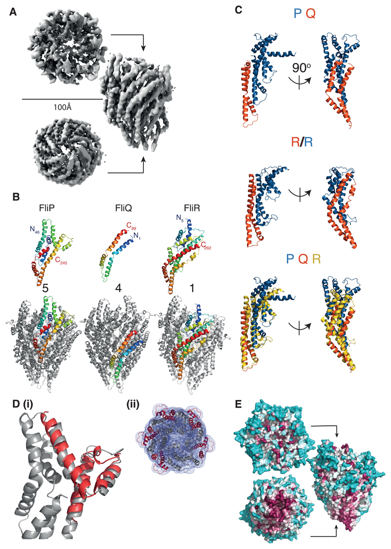
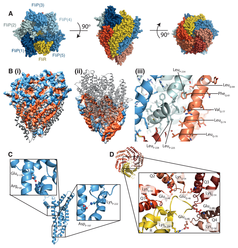
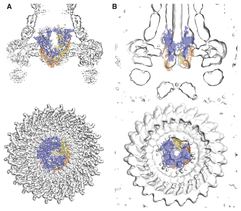
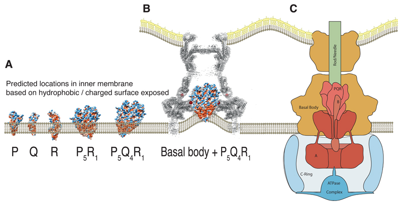
Export of proteins through type III secretion systems is critical for motility and virulence of many major bacterial pathogens. Three putative integral membrane proteins (FliP, FliQ, FliR) are suggested to form the core of an export gate in the inner membrane, but their structure, assembly and location within the final nanomachine remain unclear. Here, we present the cryoelectron microscopy structure of the Salmonella Typhimurium FliP-FliQ-FliR complex at 4.2 Å. None of the subunits adopt canonical integral membrane protein topologies, and common helix-turn-helix structural elements allow them to form a helical assembly with 5:4:1 stoichiometry. Fitting of the structure into reconstructions of intact secretion systems, combined with cross-linking, localize the export gate as a core component of the periplasmic portion of the machinery. This study thereby identifies the export gate as a key element of the secretion channel and implies that it primes the helical architecture of the components assembling downstream.


Figures

Fig. 1
Fig. 1. Stoichiometry of the PQR complexes from both Flagellar and Injectisome T3SSs revealed by native mass spectromety (nMS)
(A) Consensus topology predictions for the flagellar (FliP, FliQ & FliR) and injectisome (SctR, SctS & SctT) export gate components P, Q & R, numbered according to the S. Typhimurium flagellar sequences. The orientation of the termini of Q and R with respect to the membrane has been previously debated and they are shown in here in the same orientation as P. (B) Deconvoluted native mass spectra of complexes extracted and purified in DDM reveals a P5R1 core complex with variable numbers of Q (C) Complexes purified in the less harsh detergent LMNG contain more Q subunits, with up to the five copies of Q seen in a flagellar species complex (Table S1). Raw data are available in Supplementary Data Set 1.
Fig. 2
Fig. 2. Structure of the flagellar P5Q4R1 complex revealed at 4.2Å by cryo-electron microscopy
(A) Cryo-EM map of the P5Q4R1 complex reconstructed from 98000 particles with C1 symmetry. The complex is ~120 Å in height and the top has a diameter of ~100 Å. (B) Structures of the monomeric chains and location of an example of each within the full assembly. Each monomer is colored from blue to red at the N and C termini respectively. (C) R is a fusion of the two shorter proteins. Upper panel shows a PQ complex (P-blue;Q-orange) in two orientations with an aligned copy of R below coloured to emphasise the structural homology. The lowest panel superimposes the P (blue):Q (orange) heterodimer onto R (yellow). (D) (i)Overlay of the crystal structure (5h72 7) of the “periplasmic domain” of Thermotoga maritima FliP (red) onto one copy of FliP (grey) in the FliPQR complex. (ii) Top down view of the FliP5Q4R1 model (grey cartoon) docked in the unsharpened 4.2 Å map (grey mesh), with the five copies of the “periplasmic domain” of FliP highlighted in red. (E) Analysis of conservation using the CONSURF server reveals that the bottom of the complex is the major conserved external surface, while the region of P that adorns the outside is highly variable. The relative degree of conservation is colored dark purple for highly conserved to cyan for variable residues.
Fig. 3
Fig. 3. The P5Q4R1 complex is a right-handed helical assembly with helical parameters consistent with flagellar and injectisome assemblies.
(A) Space filling representation of the P5Q4R1 complex with R (yellow), P (shades of blue) and Q (shades of red). (B) (i) the surface of the P5R1 complex is coloured according to hydrophobicity (orange-hydrophobic; blue-charged) whilst the four copies of Q packing against a hydrophobic surface on this complex are shown as grey ribbons in (ii) the scheme is reversed and viewed from the opposite side of the complex to reveal the hydrophobic surface on the back of Q against which the P5R1 complex packs (iii) zooms in on some of the specific hydrophobic residues buried on assembly of Q. (C) Intra-molecular salt bridges within P (D) Inter-molecular salt-bridges between copies of Q and R are shown.
Fig. 4
Fig. 4. The P5Q4R1 complex is a core component of the basal body and forms a platform for assembly of the Rod.
(A) Positioning our structure within an earlier high resolution reconstruction of the injectisome basal body (grey surface and cartoon) reveals that the P5Q4R1 complex (blue cartoon) fits the un-occupied density in the center of the basal body. This region of the basal body has previously been called the “cup and socket” and sits above the proposed inner membrane location (shown as green lines). Residues on P that can be cross-linked to the basal body are highlighted by yellow spheres at the Cα position, while residues on SctC and SctJ of the basal body that cross-link to the PQR complex are shown in red. (B) In vivo photo-crosslinking studies reveal cross-links between P5Q4R1 and the inner membrane ring component SctJ in the S. Typhimurium injectisome. The residues involved are highlighted in (A). * SctJ-SctJ pBpa-independent cross-links. ** pBpa-dependent SctJ-ScJ crosslinks result in a crosslink-ladder, of which the homodimeric SctJ-SctJ crosslink is indicated by **. Representative Western blots (upper panel probed with anti-SctJ, lower panel with anti-FLAG tag), n=3. (C) Mapping of earlier cross-linking and our co-variation data (Table S3) onto P5Q4R1 reveal probable binding sites for B (FlhB or SctU) and the (inner) rod components. (D) The export gate is constricted at multiple points (i) a slab section of the entire assembly shows the levels at which the different constriction points operate (ii) views from either above or below each constriction point highlight the structural elements involved.
Fig. 5
Fig. 5. Modelling opening of the export gate.
Earlier basal body reconstructions show that the P5Q4R1 complex is closed in the absence of rod components ((A) 18) and open in their presence ((B) 21). The upper panels show slabs of the side views of these earlier maps and the lower panels shows slabs from just above the complex. In (A) the structure of the P5Q4R1 complex is fit as in Figure 4(A). An open state of the P5Q4R1 complex can be modelled (B) by superposition of models of P and R produced from co-evolution data onto our structure of the closed complex.
Fig. 6
Fig. 6. Placing the complex within the context of the full type three secretion system.
(A) Locating the isolated monomers of P, Q and R within a lipid bilayer is not trivial due to the extended nature of the hydrophobic surface and the large number of charged patches within this surface. However, the assembled object is likely to project into the periplasmic space. (B) In the absence of the other export apparatus components (A and B), earlier tomograms show the inner membrane, within the basal body, is deformed towards the region we now assign to the hydrophobic surface of Q . (C) Proposed relative locations of the five export apparatus components within the type three secretion system places the transmembrane portions of the nonameric A at the base of the P5Q4R1 complex. B is likely to form part of the helical export gate complex with the previously assumed “transmembrane” helices driving assembly and with the cytoplasmic domain hanging below a helical P5Q4R1B1 complex.

Similar articles

Cited by

References

    1. Abrusci P, McDowell MA, Lea SM, Johnson S. Building a secreting nanomachine: a structural overview of the T3SS. Curr Opin Struct Biol. 2014;25:111–117. doi: 10.1016/j.sbi.2013.11.001. - DOI - PMC - PubMed
    1. Erhardt M, Namba K, Hughes KT. Bacterial nanomachines: the flagellum and type III injectisome. Cold Spring Harb Perspect Biol. 2010;2:a000299. doi: 10.1101/cshperspect.a000299. - DOI - PMC - PubMed
    1. Buttner D. Protein export according to schedule: architecture, assembly, and regulation of type III secretion systems from plant- and animal-pathogenic bacteria. Microbiol Mol Biol Rev. 2012;76:262–310. doi: 10.1128/MMBR.05017-11. - DOI - PMC - PubMed
    1. Deng W, et al. Assembly, structure, function and regulation of type III secretion systems. Nat Rev Microbiol. 2017;15:323–337. doi: 10.1038/nrmicro.2017.20. - DOI - PubMed
    1. Macnab RM. How bacteria assemble flagella. Annu Rev Microbiol. 2003;57:77–100. doi: 10.1146/annurev.micro.57.030502.090832. - DOI - PubMed

Publication types

MeSH terms