. 2018 Jun 3:2018:8278039.
doi: 10.1155/2018/8278039. eCollection 2018.

CRP Stimulates GDF15 Expression in Endothelial Cells through p53

Affiliations

CRP Stimulates GDF15 Expression in Endothelial Cells through p53

Yoonseo Kim et al. Mediators Inflamm. .

Abstract

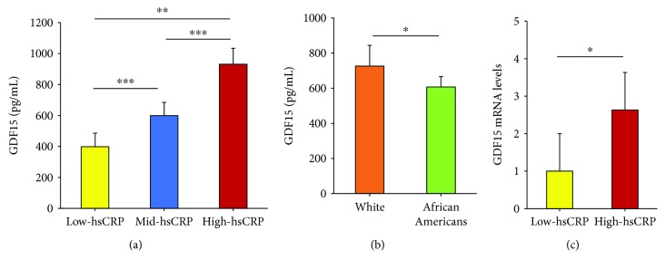
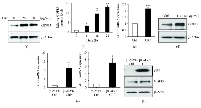
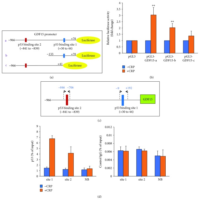
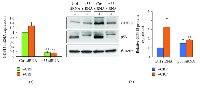
Growth differentiation factor 15 (GDF15) is a multifunctional, secreted protein that is a direct target gene of p53. GDF15 is a prospective biomarker of cardiovascular disease (CVD). C-reactive protein (CRP), like GDF15, is implicated in inflammation and an independent biomarker of CVD. However, the molecular interactions between GDF15 and CRP remain unexplored. In women, we found a significant relationship between hsCRP and GDF15 serum and mRNA levels. In vitro treatment of cultured human aortic endothelial cells (HAECs) with purified CRP or transfection of a CRP plasmid into HAECs induced GDF15 expression. Dual-luciferase reporter assays confirmed that CRP significantly increased the levels of GDF15 promoter luciferase activity, indicating that CRP induces GDF15 transcription. Chromatin immunoprecipitation (ChIP) assays confirmed that p53 was recruited to both p53 binding sites 1 and 2 in the GDF15 promoter in response to CRP. We have uncovered a linkage between CRP and GDF15, a new clue that could be important in the pathogenesis of endothelial inflammation.


Figures

Figure 1
Figure 1
Women with hsCRP have high levels of GDF15. (a, b) Serum GDF15 levels were quantified by ELISA from HANDLS participants with either low- (<3 mg/L), mid- (>3–20 mg/L), or high hsCRP (>20 mg/L) levels (n = 39/group). The ELISA assay was performed according to manufacturer's instructions and was repeated in 2 independent experiments. (c) RNA was isolated from PBMCs from HANDLS participants with either low- (<3 mg/L) or high hsCRP (>20 mg/L) levels (n = 15/group). GDF15 mRNA was quantified by RT-qPCR and normalized to HPRT1 and UBC levels. The histograms represent the mean + SEM from three independent experiments. p < 0.05, ∗∗p < 0.01, and ∗∗∗p < 0.001 by Student's t-test.
Figure 2
Figure 2
CRP upregulates GDF15 expression. (a) 18 h after CRP treatment with the indicated doses, GDF15 expression in HAECs was analyzed by immunoblotting with anti-GDF15 antibodies. β-Actin was used as a loading control. (b) After CRP treatment for the indicated time points, conditioned media was collected and GDF15 secreted levels were analyzed by ELISA. GDF15 levels were normalized to the 0 h time point for each experiment. The mean of three independent experiments is shown. (c and d) 18 h after CRP (25 μg/mL) treatment, HAECs were lysed and levels of GDF15 mRNA or protein were quantified by RT-qPCR analysis (c) and western blot analysis (d). (e) HAECs were transfected with pCMV6-control or pCMV6-CRP plasmid for 48 h. Total RNA was isolated, and mRNA levels were quantified using RT-qPCR and normalized to GAPDH. (f) Total cell lysates from the indicated transfected HAECs were analyzed by Western blotting. β-Actin was used as a loading control. The histograms represent the mean + SEM from three independent experiments. p < 0.05, ∗∗p < 0.01, and ∗∗∗p < 0.001 by Student's t-test.
Figure 3
Figure 3
CRP promotes GDF15 transcription. (a) Schematic of GDF15 promotor dual-luciferase constructs. Two p53 binding sites are indicated. (b) The indicated plasmids (1 μg) were cotransfected with 0.1 μg of TK-Renilla reporter plasmid in HeLa cells, and 24 h later, the cells were treated with CRP. After 18 h, the promoter activities were measured by luciferase activity. Transfection efficiency for luciferase activity was normalized to the Renilla luciferase activity. The results show the mean + SEM of three independent transfections. ∗∗p < 0.01 by Student's t-test. (c) Schematic of p53 binding sites and primers used for ChIP assays in the GDF15 promoter. (d) ChIP assays were performed on HAECs transfected for 24 h and treated with or without CRP for 18 h. DNA immunoprecipitated by antibodies to p53 or immunoglobulin G IgG (control) was amplified by qPCR. Each qPCR reaction was performed in triplicate, and the histogram represents the average of three independent ChIP assays + SEM.
Figure 4
Figure 4
p53 knockdown inhibits CRP-induced GDF15 expression. HAECs were transfected with either Ctrl siRNA or p53 siRNA for 24 h and treated with or without CRP for 18 h. GDF15 mRNA levels were examined by RT-qPCR (a), and protein levels were analyzed by Western blot analysis (b). The histogram represents the mean + SEM from three independent experiments. p < 0.05 and ∗∗p < 0.01 by Student's t-test.

Similar articles

Cited by

References

    1. Centers for Disease Control and Prevention. Top 10 leading causes of death: United States, 1999–2013. October 2017, https://blogs.cdc.gov/nchs-data-visualization/leading-causes-of-death/
    1. Xu J., Murphy S. L., Kochanek K. D., Bastian B. A. Deaths: final data for 2013. National Vital Statistics Reports. 2016;64(2):1–119. - PubMed
    1. Pearson T. A., Mensah G. A., Alexander R. W., et al. Markers of inflammation and cardiovascular disease: application to clinical and public health practice: a statement for healthcare professionals from the Centers for Disease Control and Prevention and the American Heart Association. Circulation. 2003;107(3):499–511. doi: 10.1161/01.CIR.0000052939.59093.45. - DOI - PubMed
    1. Bisoendial R. J., Boekholdt S. M., Vergeer M., Stroes E. S. G., Kastelein J. J. P. C-reactive protein is a mediator of cardiovascular disease. European Heart Journal. 2010;31(17):2087–2091. doi: 10.1093/eurheartj/ehq238. - DOI - PubMed
    1. Ridker P. M., Hennekens C. H., Buring J. E., Rifai N. C-reactive protein and other markers of inflammation in the prediction of cardiovascular disease in women. The New England Journal of Medicine. 2000;342(12):836–843. doi: 10.1056/NEJM200003233421202. - DOI - PubMed

LinkOut - more resources