Review
. 2018 Jun 18:9:754.
doi: 10.3389/fphys.2018.00754. eCollection 2018.

A Rising Star in Pancreatic Diseases: Pancreatic Stellate Cells

Affiliations
Review

A Rising Star in Pancreatic Diseases: Pancreatic Stellate Cells

Ran Xue et al. Front Physiol. .

Abstract

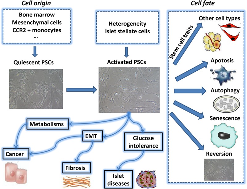
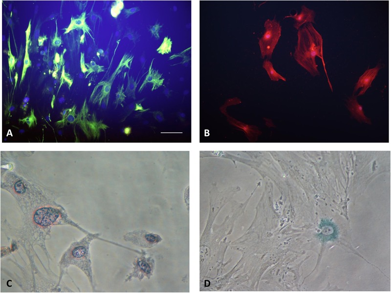
Pancreatic stellate cell (PSC) is a type of pluripotent cell located between pancreatic lobules and the surrounding area of acinars. When activated, PSC can be transformed into myofibroblast-like cell. A number of evidences suggest that activated PSC is the main source of the accumulation of extracellular matrix (ECM) protein under the pathological conditions, which lead to pancreatic fibrosis in chronic pancreatitis and pancreatic cancer. Recent studies have found that PSC also plays an important role in the endocrine cell function, islet fibrosis and diabetes. In order to provide new strategies for the treatment of pancreatic diseases, this paper systematically summarizes the recent researches about the biological behaviors of PSC, including its stem/progenitor cell characteristics, secreted exosomes, cellular senescence, epithelial mesenchymal transformation (EMT), energy metabolism and direct mechanical reprogramming.

Keywords: cellular senescence; direct mechanical reprogramming; energy metabolism; epithelial mesenchymal transformation; exosomes; pancreatic stellate cell; stem/progenitor cell characteristics.


Figures

FIGURE 1
FIGURE 1
The above summarizes the major topics of biological behavior of pancreatic stellate cell which are covered during this review period. PSC, pancreatic stellate cell; EMT, epithelial-mesenchymal transition.
FIGURE 2
FIGURE 2
(A) Quiescent PSCs stain positively for desmin by IF. (Hoechst 33258 staining, and Alexa Fluor 488 staining for desmin, original magnification, ×200). (B) Activated PSCs stain positively for α-SMA by IF (Hoechst 33258 staining, and Alexa Fluor 594 staining for α-SMA, original magnification, ×200). (C) Quiescent PSCs have an angular appearance, contained lipid droplets by oil red O staining. (Original magnification, ×400). (D) Senescence is shown in PSCs with cytoplasmic blue staining of SA-β-gal. (Immunocytochemistry; original magnification, ×400) (Xue et al., 2017b).

Similar articles

Cited by

References

    1. Al-Assar O., Demiciorglu F., Lunardi S., Gaspar-Carvalho M. M., McKenna W. G., Muschel R. M., et al. (2014). Contextual regulation of pancreatic cancer stem cell phenotype and radioresistance by pancreatic stellate cells. Radiother. Oncol. 111 243–251. 10.1016/j.radonc.2014.03.014 - DOI - PubMed
    1. Apte M. V., Haber P. S., Applegate T. L., Norton I. D., McCaughan G. W., Korsten M. A., et al. (1998). Periacinar stellate shaped cells in rat pancreas: identification, isolation, and culture. Gut. 43 128–133. 10.1136/gut.43.1.128 - DOI - PMC - PubMed
    1. Apte M. V., Wilson J. S., Lugea A., Pandol S. J. (2013a). A starring role for stellate cells in the pancreatic cancer microenvironment. Gastroenterology 144 1210–1219. 10.1053/j.gastro.2012.11.037 - DOI - PMC - PubMed
    1. Apte M. V., Yang L., Phillips P. A., Xu Z., Kaplan W., Cowley M., et al. (2013b). Extracellular matrix composition significantly influences pancreatic stellate cell gene expression pattern: role of transgelin in PSC function. Am. J. Physiol. Gastrointest. Liver Physiol. 305 G408–G417. 10.1152/ajpgi.00016.2013 - DOI - PubMed
    1. Asahina K., Zhou B., Pu W. T., Tsukamoto H. (2011). Septum transversum-derived mesothelium gives rise to hepatic stellate cells and perivascularmesenchymal cells in developing mouse liver. Hepatology 53 983–995. 10.1002/hep.24119 - DOI - PMC - PubMed

LinkOut - more resources