. 2018 Jun 18:9:1358.
doi: 10.3389/fimmu.2018.01358. eCollection 2018.

Hodgkin Lymphoma-Derived Extracellular Vesicles Change the Secretome of Fibroblasts Toward a CAF Phenotype

Affiliations

Hodgkin Lymphoma-Derived Extracellular Vesicles Change the Secretome of Fibroblasts Toward a CAF Phenotype

Bastian Dörsam et al. Front Immunol. .

Abstract

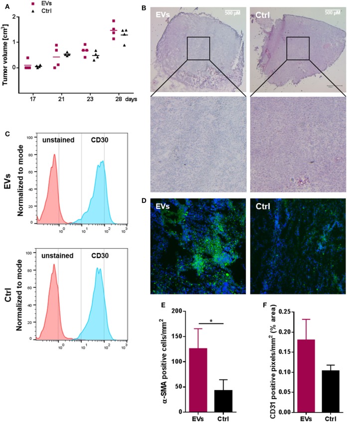
Secretion of extracellular vesicles (EVs) is a ubiquitous mechanism of intercellular communication based on the exchange of effector molecules, such as growth factors, cytokines, and nucleic acids. Recent studies identified tumor-derived EVs as central players in tumor progression and the establishment of the tumor microenvironment (TME). However, studies on EVs from classical Hodgkin lymphoma (cHL) are limited. The growth of malignant Hodgkin and Reed-Sternberg (HRS) cells depends on the TME, which is actively shaped by a complex interaction of HRS cells and stromal cells, such as fibroblasts and immune cells. HRS cells secrete cytokines and angiogenic factors thus recruiting and inducing the proliferation of surrounding cells to finally deploy an immunosuppressive TME. In this study, we aimed to investigate the role of tumor cell-derived EVs within this complex scenario. We observed that EVs collected from Hodgkin lymphoma (HL) cells were internalized by fibroblasts and triggered their migration capacity. EV-treated fibroblasts were characterized by an inflammatory phenotype and an upregulation of alpha-smooth muscle actin (α-SMA), a marker of cancer-associated fibroblasts. Analysis of the secretome of EV-treated fibroblast revealed an enhanced release of pro-inflammatory cytokines (e.g., IL-1α, IL-6, and TNF-α), growth factors (G-CSF and GM-CSF), and pro-angiogenic factors such as VEGF. These soluble factors are known to promote HL progression. In line, ingenuity pathway analysis identified inflammatory pathways, including TNF-α/NF-κB-signaling, as key factors directing the EV-dependent phenotype changes in fibroblasts. Confirming the in vitro data, we demonstrated that EVs promote α-SMA expression in fibroblasts and the expression of proangiogenic factors using a xenograft HL model. Collectively, we demonstrate that HL EVs alter the phenotype of fibroblasts to support tumor growth, and thus shed light on the role of EVs for the establishment of the tumor-promoting TME in HL.

Keywords: Hodgkin lymphoma; NF-κB-signaling; cancer-associated fibroblasts; extracellular vesicles; tumor microenvironment.


Figures

Figure 1
Figure 1
Characterization and internalization of tumor-derived extracellular vesicles (EVs) by fibroblast cells in vitro. (A) Size distribution of fibroblast EVs (performed once) or Hodgkin lymphoma (HL) EVs (representative experiment, n = 3), measured by nanoparticle tracking analysis. (B) Electron microscopic image of purified HL EVs (n = 1). (C) Western blot analysis of exosome markers on HL-derived EVs. Cell lysate of HL cells served as control. Presented is one of three independent experiments. (D) Flow cytometry of beads coupled HL EVs which was collected from unstained cells (Ctrl) or from DiO-stained cells (0.25 or 1 µM DiO, n = 1). Normalization to mode: events are normalized in a scale with 100 being maximum (FlowJo v10). (E) Internalization of DiO-labeled HL EVs by fibroblasts assessed via flow cytometry at different time points as indicated. Fibroblast cells were incubated with 100 µg DiO+-EVs or DiO-negative EVs as negative control in the presence of EV-depleted medium. Statistical significance was calculated with one-way ANOVA and Tukey’s multiple comparisons test (mean + SEM of three biological replicates; *p ≥ 0.05). (F) Visualization of EV-uptake into fibroblasts via immunofluorescence: cytoplasm was stained with Cell Mask Deep Red (red), nuclei with Hoechst and HL-derived EVs with DiO (green). Upper picture shows a recipient cell with internalized HL EVs. Bottom picture depicts a three-dimensional view.
Figure 2
Figure 2
Extracellular vesicles (EVs) promote migration of Hodgkin lymphoma (HL) cells and HL EVs can alter the phenotype of fibroblast cells. (A) Transwell migration assay (8 µm pores): HL cells were exposed to supernatant from fibroblast cells or EVs for 26 h. Statistical significance was calculated with one-way ANOVA and Tukey’s multiple comparisons test (presented is mean + SEM, four biological replicates; ***p ≥ 0.001; ****p ≥ 0.0001). (B) Scratch assay: 24 h wound closure of fibroblast cells exposed to HL EVs (green) or medium (black). Student’s t-test was performed to check for significant differences between both treatments (mean ± SEM, n = 3; *p ≥ 0.05; **p ≥ 0.01). (C) Chemotaxis assay: migration of labeled fibroblast cells through Neuroprobe ChemoTx plates (5 µm pores) exposed to medium, 40 or 150 µg/ml HL EVs. Statistical assessment was performed with one-way ANOVA and Tukey’s multiple comparisons test (data are presented as mean + SEM, n = 4; *p ≥ 0.05; **p ≥ 0.01). (D) Exemplary bright field and fluorescence pictures of a scratch assay after 24 h exposure of fibroblasts to 100 µg/ml (≈1 × 109 EVs/ml) HL EVs or medium. Nuclei stained with DAPI (blue), fibroblast cells with α-SMA (green). This experiment was performed in three independent replicates. (E) Human 64-Plex chemokine array: quantification of chemokines/cytokines after 24 h in the supernatant of fibroblasts in presence or absence of HL EVs. Not detected cytokines/chemokines are depicted in italic letters; abundant factors are in bold; enhanced expression is highlighted in green, whereas red boxes indicate lower expression after exposure to HL EVs. Statistical differences in abundance of chemokines/cytokine were determined using Student’s t-test (three independent replicates).
Figure 3
Figure 3
Hodgkin lymphoma (HL) xenograft model. 1 × 107 HDFn + 1 × 107 KM-H2 cells were subcutaneously transplanted into NOD scid gamma mice (age 141 days). Treatment group (n = 4 animals) received an i.v. injection of HL extracellular vesicles (EVs) administered via the tail vein at day 22, 25, 27, and 28, animals of the control group (n = 4 animals) were injected with PBS. Necropsy was performed on day 30. (A) Tumor growth in EV-treated and control animals; tumor volume was assessed as tumors were detectable (n = 4 animals per group). (B) Representative hematoxylin and eosin stainings of tumor tissue cryo-sections (25× magnification) from EV-treated (EVs) or control (Ctrl) animals (four animals per group were analyzed). (C) Abundance of CD30 on cells of four resected tumors analyzed by flow cytometry. (D) DiO-positive tumor cells after internalization of labeled HL EVs (n = 4). (E) Number of α-SMA positive cells and (F) cells expressing the vascularization marker CD31 in tumor sections of both groups assessed microscopically and quantified using ImageJ software (http://rsb.info.nih.gov/ij) (Student’s t-test of n ≥ 3 samples presented as mean + SEM; *p ≥ 0.05).
Figure 4
Figure 4
Interaction of fibroblasts and Hodgkin lymphoma (HL) cells in the tumor microenvironment. Communication of the few malignant cells in HL with fibroblasts is crucial for survival of the tumor cells. Due to the spatial distance between the cells, direct cell–cell interaction is not the first-line communication mechanism. Interaction of both cell types is facilitated by soluble and vesicular factors, e.g., chemokines/cytokines, which enhance motility and migration of cells. Further, HL extracellular vesicles shape the phenotype of fibroblasts, skewing the cells to a cancer-associated cell state concomitant with alteration of their secretome. Our results suggest a strong involvement of the TNF-α/NF-κB axis in this process.

Similar articles

Cited by

References

    1. Hodgkin T. On some morbid appearances of the absorbent glands and spleen. Med Chir Trans (1832) 17:68–114.10.1177/095952873201700106 - DOI - PMC - PubMed
    1. DeVita VT, Costa J. Toward a personalized treatment of Hodgkin’s disease. N Engl J Med (2010) 362:942–3.10.1056/NEJMe0912481 - DOI - PubMed
    1. Küppers R, Engert A, Hansmann M-L. Hodgkin lymphoma. J Clin Invest (2012) 122:3439–47.10.1172/JCI61245 - DOI - PMC - PubMed
    1. Aldinucci D, Gloghini A, Pinto A, de Filippi R, Carbone A. The classical Hodgkin’s lymphoma microenvironment and its role in promoting tumour growth and immune escape. J Pathol (2010) 221:248–63.10.1002/path.2711 - DOI - PubMed
    1. Tzankov A, Zimpfer A, Pehrs A-C, Lugli A, Went P, Maurer R, et al. Expression of B-cell markers in classical Hodgkin lymphoma: a tissue microarray analysis of 330 cases. Mod Pathol (2003) 16:1141–7.10.1097/01.MP.0000093627.51090.3F - DOI - PubMed

LinkOut - more resources