

Transcriptome Analysis of Bronchoalveolar Lavage Fluid From Children With Mycoplasma pneumoniae Pneumonia Reveals Natural Killer and T Cell-Proliferation Responses
- PMID: 29967623
- PMCID: PMC6015898
- DOI: 10.3389/fimmu.2018.01403
Transcriptome Analysis of Bronchoalveolar Lavage Fluid From Children With Mycoplasma pneumoniae Pneumonia Reveals Natural Killer and T Cell-Proliferation Responses
Abstract
Background: Mycoplasma pneumoniae pneumonia (MPP) is one of the most common community-acquired pneumonia; this study is to explore the immune-pathogenesis of children MPP.
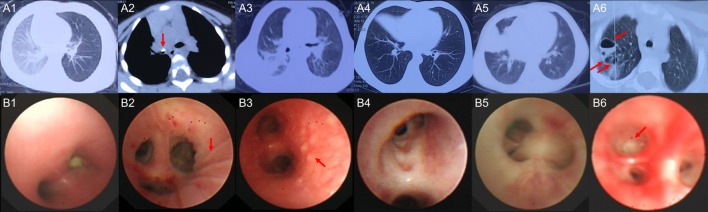
Methods: Next-generation transcriptome sequencing was performed on the bronchoalveolar lavage fluid cells from six children with MPP and three children with foreign body aspiration as control. Some of the results had been validated by quantitative real-time PCR in an expanded group of children.
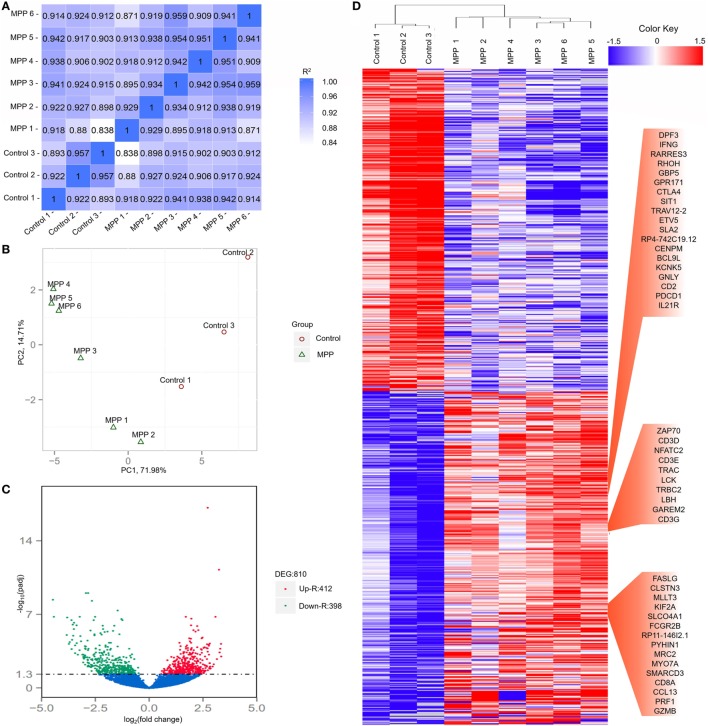
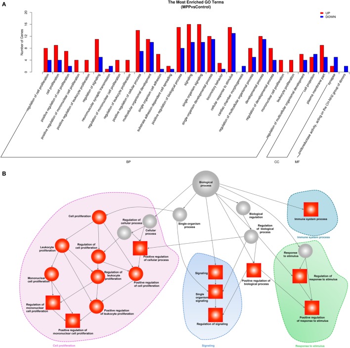
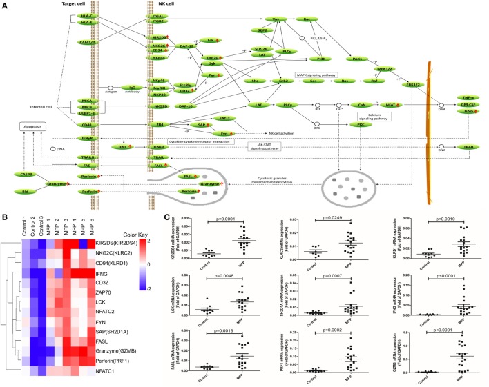
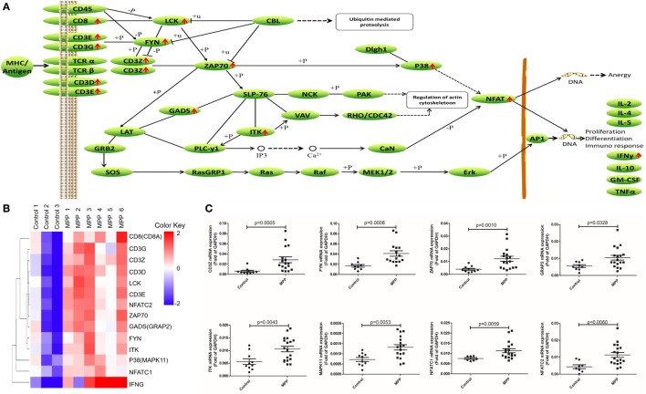
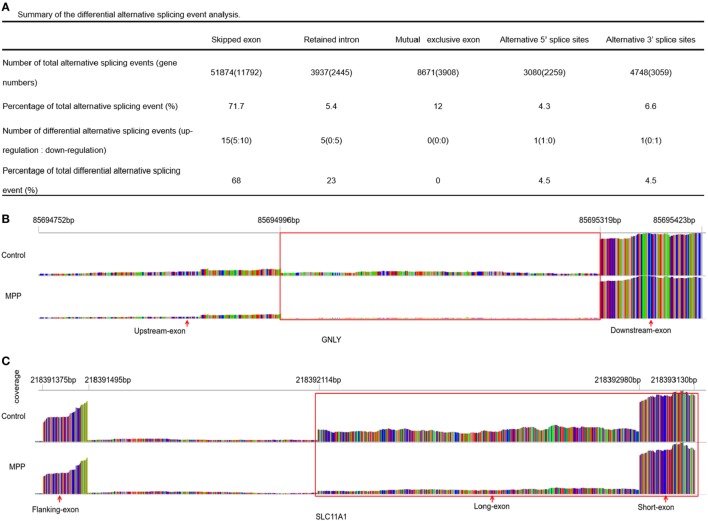
Results: Results revealed 810 differentially expressed genes in MPP group comparing to control group, of which 412 genes including RLTPR, CARD11 and RASAL3 were upregulated. These upregulated genes were mainly enriched in mononuclear cell proliferation and signaling biological processes. Kyoto encyclopedia of genes and genomes pathway analysis revealed that hematopoietic cell linage pathway, natural killer cell-mediated cytotoxicity pathway, and T cell receptor signaling pathway were significantly upregulated in MPP children. In addition, significant alternative splicing events were found in GNLY and SLC11A1 genes, which may cause the differential expressions of these genes.
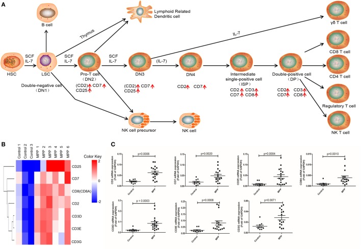
Conclusion: Our results suggest that NK and CD8+ T cells are over activated and proliferated in MPP children; the upregulated IFNγ, PRF1, GZMB, FASL, and GNLY may play important roles in the pathogenesis of children MPP.
Keywords: CD8+ T cells; Mycoplasma pneumoniae pneumonia; bronchoalveolar lavage fluid; children; interferon gamma; natural killer cells.
Figures
Similar articles
-
Transcriptome analysis of bronchoalveolar lavage fluid from children with severe Mycoplasma pneumoniae pneumonia reveals novel gene expression and immunodeficiency.Hum Genomics. 2017 Mar 16;11(1):4. doi: 10.1186/s40246-017-0101-y. Hum Genomics. 2017. PMID: 28302172 Free PMC article.
-
Proteomic characteristics of bronchoalveolar lavage fluid in children with mild and severe Mycoplasma pneumoniae pneumonia.Front Microbiol. 2025 May 19;16:1595521. doi: 10.3389/fmicb.2025.1595521. eCollection 2025. Front Microbiol. 2025. PMID: 40458711 Free PMC article.
-
Immune response plays a role in Mycoplasma pneumoniae pneumonia.Front Immunol. 2023 May 26;14:1189647. doi: 10.3389/fimmu.2023.1189647. eCollection 2023. Front Immunol. 2023. PMID: 37304280 Free PMC article.
-
Serum Tumor Necrosis Factor-α and Interferon-γ Levels in Pediatric Mycoplasma pneumoniae Pneumonia: A Systematic Review and Meta-Analysis.Can Respir J. 2018 Sep 10;2018:8354892. doi: 10.1155/2018/8354892. eCollection 2018. Can Respir J. 2018. PMID: 30275916 Free PMC article.
-
Rational stepwise approach for Mycoplasma pneumoniae pneumonia in children.J Microbiol Immunol Infect. 2021 Aug;54(4):557-565. doi: 10.1016/j.jmii.2020.10.002. Epub 2020 Oct 17. J Microbiol Immunol Infect. 2021. PMID: 33268306 Review.
Cited by
-
Temperature impacts the bovine ex vivo immune response towards Mycoplasmopsis bovis.Vet Res. 2024 Feb 13;55(1):18. doi: 10.1186/s13567-024-01272-3. Vet Res. 2024. PMID: 38351086 Free PMC article.
-
High co-expression of TNF-α and CARDS toxin is a good predictor for refractory Mycoplasma pneumoniae pneumonia.Mol Med. 2019 Aug 9;25(1):38. doi: 10.1186/s10020-019-0105-2. Mol Med. 2019. PMID: 31399022 Free PMC article.
-
Effects of bronchoalveolar lavage on Mycoplasma Pneumoniae pneumonia: A propensity score matched-cohort study.Front Pediatr. 2023 Jan 4;10:1066640. doi: 10.3389/fped.2022.1066640. eCollection 2022. Front Pediatr. 2023. PMID: 36683805 Free PMC article.
-
LncRNA MALAT1 Affects Mycoplasma pneumoniae Pneumonia via NF-κB Regulation.Front Cell Dev Biol. 2020 Oct 2;8:563693. doi: 10.3389/fcell.2020.563693. eCollection 2020. Front Cell Dev Biol. 2020. PMID: 33134293 Free PMC article.
-
Attenuated lncRNA NKILA Enhances the Secretory Function of Airway Epithelial Cells Stimulated by Mycoplasma pneumoniae via NF-κB.Biomed Res Int. 2021 Mar 26;2021:6656298. doi: 10.1155/2021/6656298. eCollection 2021. Biomed Res Int. 2021. PMID: 33855076 Free PMC article.
References
LinkOut - more resources
Full Text Sources
Other Literature Sources
Research Materials