

The Bioactive Substance Secreted by MSC Retards Mouse Aortic Vascular Smooth Muscle Cells Calcification
- PMID: 29967775
- PMCID: PMC6008760
- DOI: 10.1155/2018/6053567
The Bioactive Substance Secreted by MSC Retards Mouse Aortic Vascular Smooth Muscle Cells Calcification
Abstract
Background: Vascular calcification, which is associated with low-level chronic inflammation, is a complication that occurs during aging, atherosclerosis, chronic kidney disease, diabetes mellitus, and hyperlipaemia. In this study, we used conditioned media from mesenchymal stem cells (MSC-CM), a source of autologous cytokines, to test the hypothesis that MSC-CM inhibits vascular smooth muscle cell (VSMC) calcification by suppressing inflammation and apoptosis.
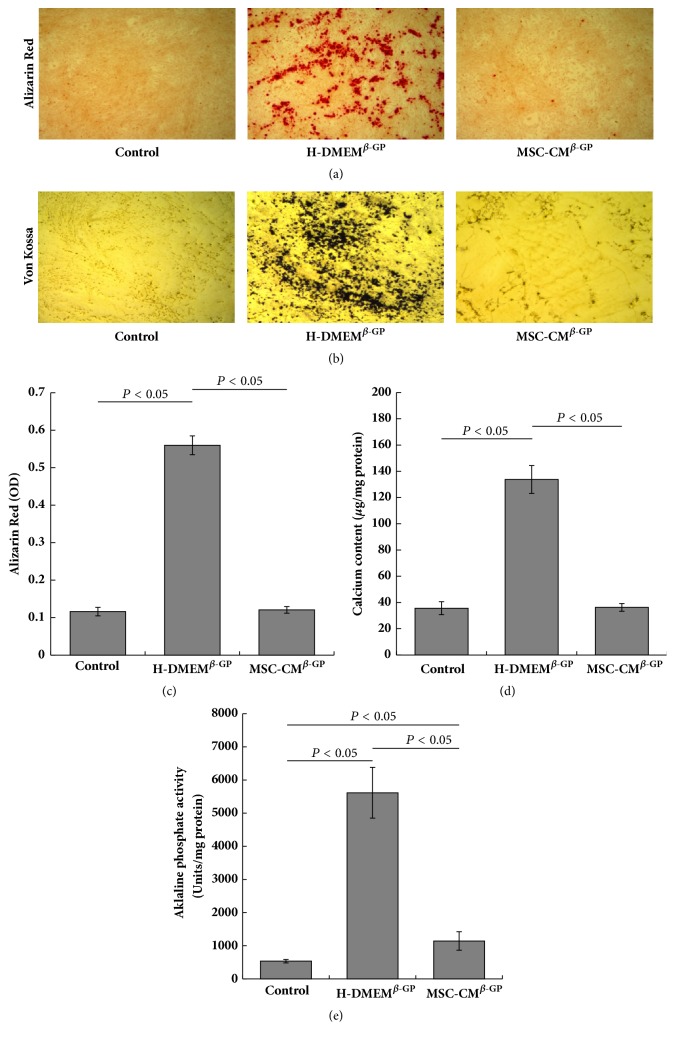
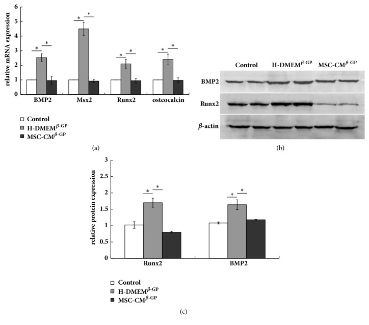
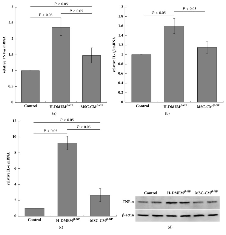
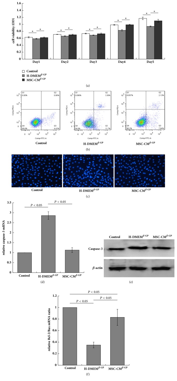
Methods: VSMCs were treated with β-glycerophosphate (β-GP) to induce calcification and MSC-CM was used as a treatment. Calcium deposition was evaluated using alizarin red and von Kossa staining after a 7-day induction period. Intracellular calcium contents were measured via the o-cresolphthalein complexone method, and alkaline phosphatase (ALP) activity was determined using the para-nitrophenyl phosphate method. The expressions of specific-osteogenic markers, inflammatory cytokines, and apoptosis-associated genes/proteins were examined by real-time polymerase chain reaction or western blotting.
Results: MSC-CM inhibited β-GP-induced calcium deposition in VSMCs and decreased intracellular calcium content and ALP activity. Additionally, MSC-CM suppressed the β-GP-induced increases in BMP2, Msx2, Runx2, and osteocalcin expression. Additionally, MSC-CM decreased the expression of TNF-α, IL-1β, and IL-6 in VSMC. MSC-CM also partly blocked β-GP-induced VSMC apoptosis, which was associated with an increase in the Bcl-2/Bax expression ratio and a decrease in caspase-3 expression.
Conclusion: Our study results suggest that MSC-CM can inhibit VSMC calcification. This suggests a potential novel clinical application for MSCs in the treatment of vascular calcification and associated diseases.
Figures
References
-
- Deuell K. A., Callegari A., Giachelli C. M., Rosenfeld M. E., Scatena M. RANKL enhances macrophage paracrine pro-calcific activity in high phosphate-treated smooth muscle cells: Dependence on IL-6 and TNF-α. Journal of Vascular Research. 2012;49(6):510–521. doi: 10.1159/000341216. - DOI - PMC - PubMed
-
- Proudfoot D., Skepper J. N., Hegyi L., Bennett M. R., Shanahan C. M., Weissberg P. L. Apoptosis regulates human vascular calcification in vitro: evidence for initiation of vascular calcification by apoptotic bodies. Circulation Research. 2000;87(11):1055–1062. doi: 10.1161/01.res.87.11.1055. - DOI - PubMed
MeSH terms
Substances
LinkOut - more resources
Full Text Sources
Other Literature Sources
Research Materials
