. 2018 Aug;136(2):181-210.
doi: 10.1007/s00401-018-1879-y. Epub 2018 Jul 2.

Practical implementation of DNA methylation and copy-number-based CNS tumor diagnostics: the Heidelberg experience

Affiliations

Practical implementation of DNA methylation and copy-number-based CNS tumor diagnostics: the Heidelberg experience

David Capper et al. Acta Neuropathol. 2018 Aug.

Abstract

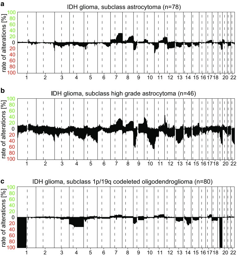
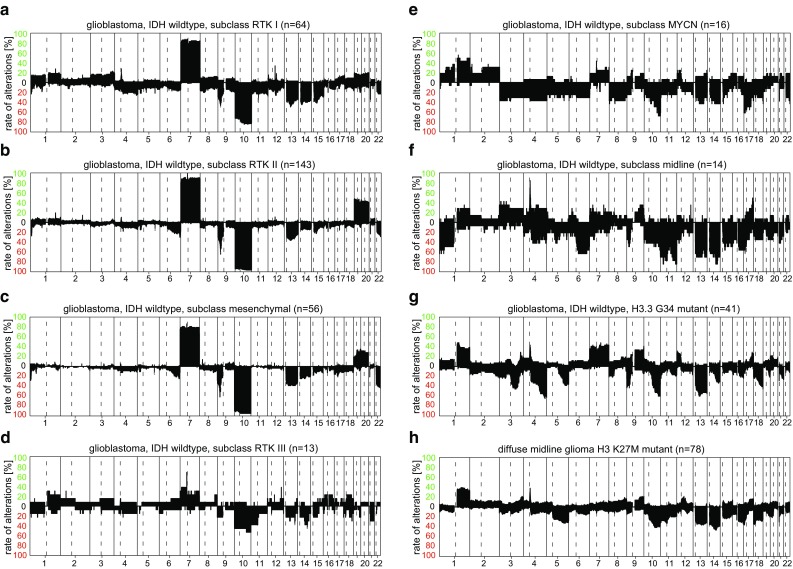
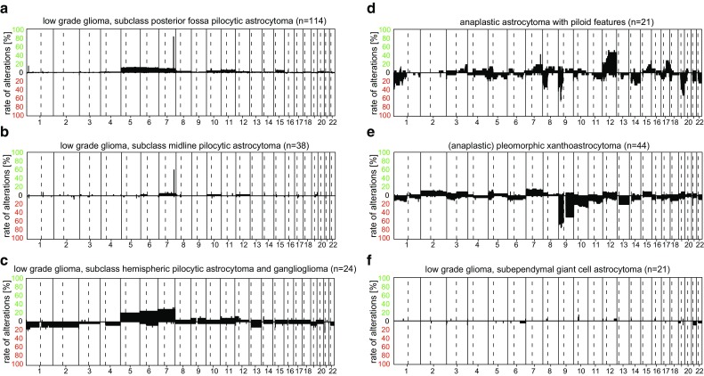
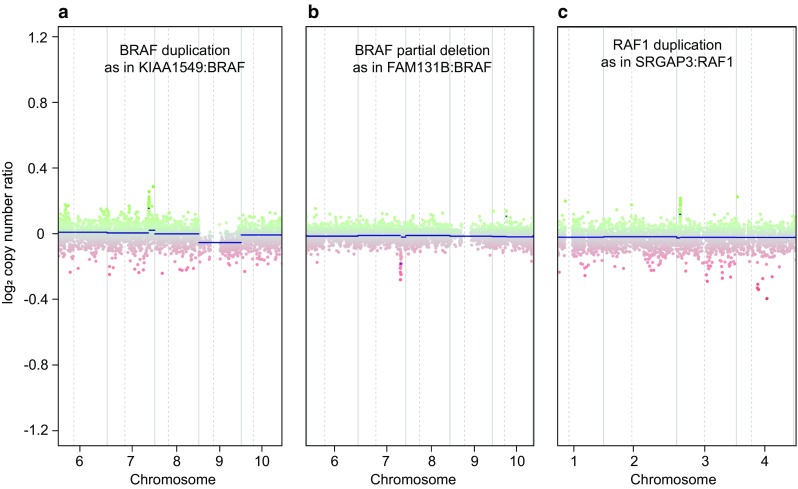
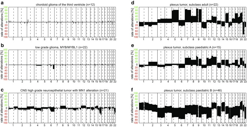
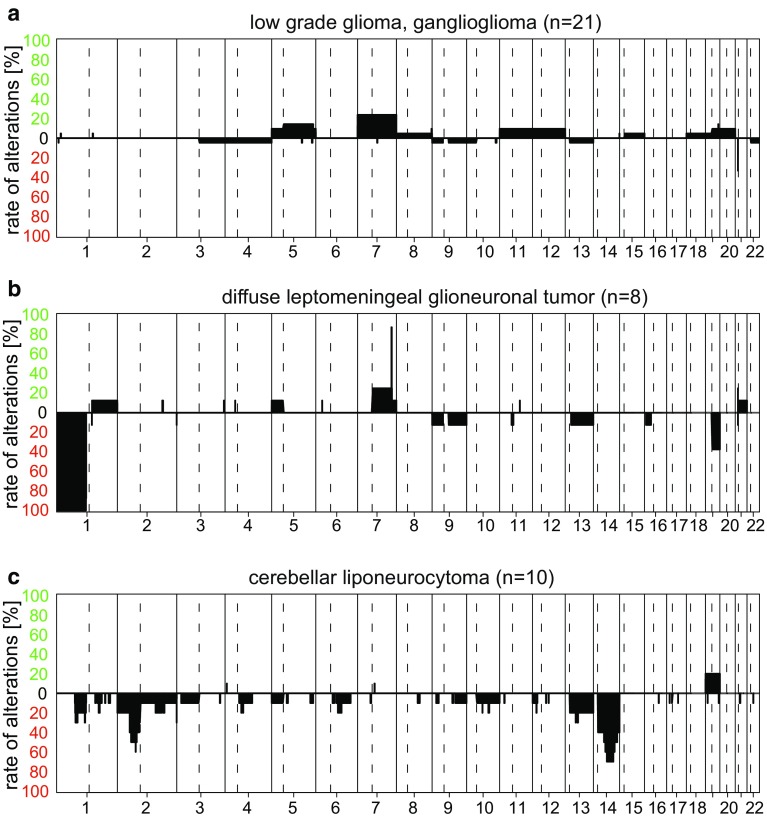
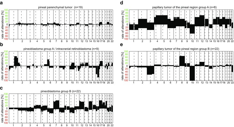
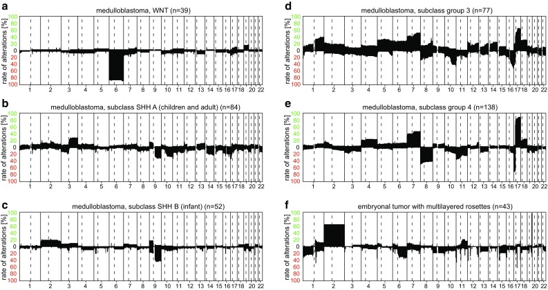
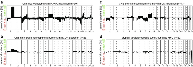
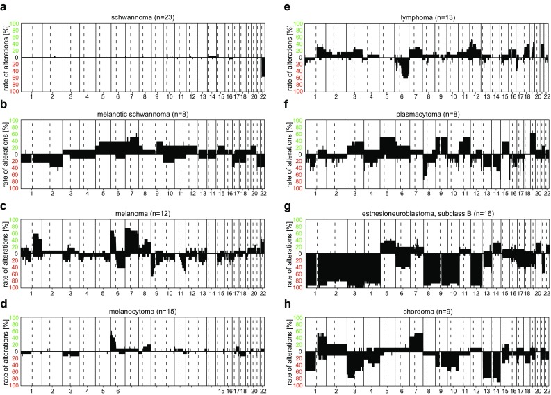
Recently, we described a machine learning approach for classification of central nervous system tumors based on the analysis of genome-wide DNA methylation patterns [6]. Here, we report on DNA methylation-based central nervous system (CNS) tumor diagnostics conducted in our institution between the years 2015 and 2018. In this period, more than 1000 tumors from the neurosurgical departments in Heidelberg and Mannheim and more than 1000 tumors referred from external institutions were subjected to DNA methylation analysis for diagnostic purposes. We describe our current approach to the integrated diagnosis of CNS tumors with a focus on constellations with conflicts between morphological and molecular genetic findings. We further describe the benefit of integrating DNA copy-number alterations into diagnostic considerations and provide a catalog of copy-number changes for individual DNA methylation classes. We also point to several pitfalls accompanying the diagnostic implementation of DNA methylation profiling and give practical suggestions for recurring diagnostic scenarios.

Keywords: Copy-number variation; DNA methylation; EPIC array; Tumor classification.


Figures

Fig. 1
Fig. 1
CNV plots calculated from DNA methylation array data. a Example of a typical CNV plot of a glioblastoma, IDH wt (subclass receptor tyrosine kinase (RTK) II). Depiction of Chromosome 1–22 with the p-arm (left) and the q-arm (right) separated by a dotted line. Gains/amplifications represent positive, losses negative deviations from the baseline. The probes of the array are combined in 8000 bins (green or red dots). For assessment of relevant deviations from the baseline, we, in general, only consider deviations of the dark blue line that represents an average over several dots and not individual colored dots. This case shows a gain of chromosomes Chr.7, 19, and 20 likely representing trisomies and several sub-chromosomal losses of one chromosomal copy (the largest of Chr.10q and several smaller ones, e.g., two on Chr.1p and one on Chr.4q). In addition, the case shows an amplification of EGFR (highly focal shifting of the blue line accompanied by a row of single green dots including the EGFR locus, usually above a log2 value of 0.4) and a homozygous deletion of CDKN2A/B (focal shifting of the blue line accompanied by a row or single red dots including the CDKN2A/B locus usually below a log2 value of 0.4). Besides EGFR and CDKN2A/B, 27 other gene regions are highlighted by default by the gene name and a blue dot for easier identification of possible copy-number alterations. The highlighting does not indicate relevant changes per se. Low-tumor cell content or subclonal alterations may reduce the amplitude of blue line deviation, and thus, a general cut-off value for what deviation is a definite chromosomal change cannot be provided. b Example of a CNV plot of an RELA-fused ependymoma with artificial noise caused by an unsuccessful bisulfite conversion. CNV information should not be extracted from plots of this quality. c Reanalysis of the same case as in b) without technical issues now demonstrating a crisp CNV plot that can be clearly interpreted. In line with the Classifier result, it shows changes compatible with an RELA- fused ependymoma (see Fig. 7g for comparison). d Unusual CNV plot of a non-classifiable case with multiple whole chromosomal gains and losses (likely representing a carcinoma metastasis) and signs of cross contamination with DNA from the sample in e (glioblastoma, IDH Wt). The identical three amplifications are found in both cases. This is the result of minimal amounts of DNA exchange from e to d during array preparation
Fig. 2
Fig. 2
Summary CNV plots of IDH mutant diffuse astrocytic and oligodendroglial tumors. a IDH glioma, subclass astrocytoma; b IDH glioma, subclass high-grade astrocytoma; c IDH glioma, subclass 1p/19q codeleted oligodendroglioma
Fig. 3
Fig. 3
Summary CNV plots of seven IDH wt glioblastoma methylation classes (ag) and diffuse midline glioma, H3 K27M mutant (h)
Fig. 4
Fig. 4
Example of cases with conflicting pathological and molecular results. a H&E of an IDH wt, moderately cell dense, moderately pleomorphic diffuse glioma diagnosed as diffuse astrocytoma, IDH wt (WHO grade II). b Methylation class and CNV plot correspond to a glioblastoma, IDH wt. The integrated diagnosis was also glioblastoma, IDH wt. Many of these cases are from small biopsies. c H&E of a pleomorphic necrotizing tumor with the pathological diagnosis of glioblastoma, IDH wt. d Methylation class corresponds to an (anaplastic) pleomorphic xanthoastrocytoma; the copy-number profile would be compatible with both diagnoses (and is not highly characteristic for either). A BRAF V600 mutation was not present. This conflict could not be resolved and the case was diagnosed as malignant glioma, histologically glioblastoma, methylation profile (anaplastic) PXA, NEC for the time being
Fig. 5
Fig. 5
Summary CNV plots of “other astrocytic tumors” including subclasses of pilocytic astrocytoma (ac), anaplastic astrocytoma with piloid features (d), (anaplastic) pleomorphic xanthoastrocytoma (e), and subependymal giant cell astrocytoma (f)
Fig. 6
Fig. 6
Examples of three pilocytic astrocytomas with different types of MAPK alterations visible on the CNV plots. a Shows the by far most frequent focal low-level gain on Chr.7q representing a 7q34 tandem duplication of BRAF as part of the KIAA1549:BRAF gene fusion; b represents a rare fusion event with a focal loss of representing FAM131B:BRAF gene fusion; c represents a focal gain involving RAF1 as part of the SRGAP3:RAF1 gene fusion
Fig. 7
Fig. 7
Summary CNV plots of ependymal and subependymal tumors (ah)
Fig. 8
Fig. 8
Summary CNV plots of a selection of “other gliomas” (ac) and choroid plexus tumors (df)
Fig. 9
Fig. 9
Summary CNV plots of a selection of Neuronal and mixed neuronal–glial tumors (ac)
Fig. 10
Fig. 10
Example of cases with conflicting pathological and molecular results. a H&E of a tumor composed of relatively monomorphous, indistinct cells pathologically interpreted as diffuse astrocytoma, IDH wt. b By methylation profiling, the tumor corresponds to a ganglioglioma. The copy-number profile is flat and would also fit to the latter (but is also not specific for this). BRAF sequencing revealed a V600E mutation. The case was interpreted as ganglioglioma in the integrated diagnosis. c H&E of a supratentorial embryonal tumor, NEC with a Ki67 proliferation fraction of approximately 20% (not shown) that by methylation profiling, d corresponds to a diffuse leptomeningeal glioneuronal tumor. A BRAF duplication was not present. Anaplastic transition of diffuse leptomeningeal glioneuronal tumor has been documented [38]. Likely, the histological spectrum of tumors with the molecular profile of diffuse leptomeningeal glioneuronal tumors is even wider [8] and the way to best categorize such cases remains to be established
Fig. 11
Fig. 11
Summary CNV plots of pineal region tumors (ae)
Fig. 12
Fig. 12
Example of a case with conflicting pathological and molecular results. H&E of tumor of a pediatric patient showing high resemblance to a papillary tumor of the pineal region, but that is located in the 4th ventricle and had no contact to the pineal region. The DNA methylation profile also corresponded to papillary tumor of the pineal region and the CNV profile would also be compatible with this. It is not clear whether this represents an ectopic papillary tumor of the pineal region or a distinct so far not defined tumor class
Fig. 13
Fig. 13
Summary CNV plots of medulloblastoma sub-groups (ae) and embryonal tumor with multilayered rosettes (f)
Fig. 14
Fig. 14
Summary CNV plots of a selection of embryonal tumors (ad)
Fig. 15
Fig. 15
Summary CNV plots of a selection of tumors of the cranial and paraspinal nerves (ab), melanocytic tumors (cd), large B-cell lymphoma (e), and plasmacytoma (f), and selected other intracranial tumors (gh)

Similar articles

Cited by

References

    1. Bady P, Delorenzi M, Hegi ME. Sensitivity analysis of the MGMT-STP27 model and impact of genetic and epigenetic context to predict the MGMT methylation status in gliomas and other tumors. J Mol Diagn. 2016;18:350–361. doi: 10.1016/j.jmoldx.2015.11.009. - DOI - PubMed
    1. Bady P, Sciuscio D, Diserens AC, et al. MGMT methylation analysis of glioblastoma on the Infinium methylation BeadChip identifies two distinct CpG regions associated with gene silencing and outcome, yielding a prediction model for comparisons across datasets, tumor grades, and CIMP-status. Acta Neuropathol. 2012;124:547–560. doi: 10.1007/s00401-012-1016-2. - DOI - PMC - PubMed
    1. Brastianos PK, Taylor-Weiner A, Manley PE, et al. Exome sequencing identifies BRAF mutations in papillary craniopharyngiomas. Nat Genet. 2014;46:161–165. doi: 10.1038/ng.2868. - DOI - PMC - PubMed
    1. Cancer Genome Atlas Research N. Brat DJ, Verhaak RG, et al. Comprehensive, integrative genomic analysis of diffuse lower-grade gliomas. N Engl J Med. 2015;372:2481–2498. doi: 10.1056/NEJMoa1402121. - DOI - PMC - PubMed
    1. Capper D, Engel NW, Stichel D et al (2018) DNA methylation-based reclassification of olfactory neuroblastoma. Acta Neuropathol. 10.1007/s00401-018-1854-7 - PubMed

MeSH terms