. 2018 Oct;183(5):751-764.
doi: 10.1007/s11046-018-0277-2. Epub 2018 Jul 2.

The Oomycete Pythium oligandrum Can Suppress and Kill the Causative Agents of Dermatophytoses

Affiliations

The Oomycete Pythium oligandrum Can Suppress and Kill the Causative Agents of Dermatophytoses

Alena Gabrielová et al. Mycopathologia. 2018 Oct.

Abstract

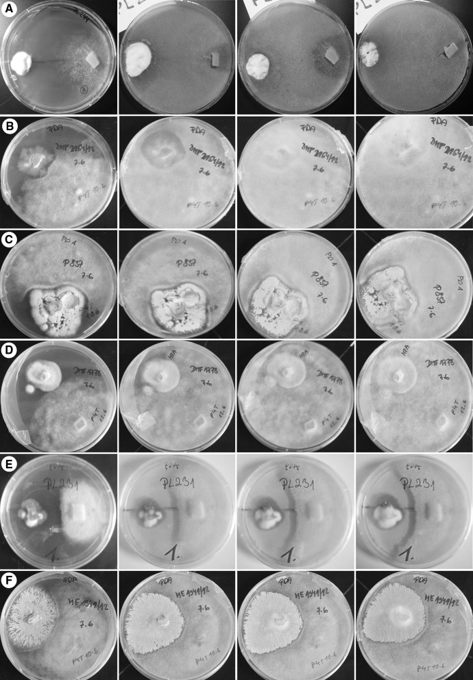
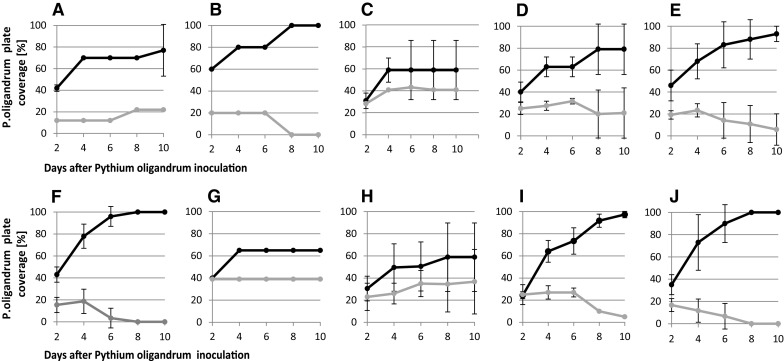
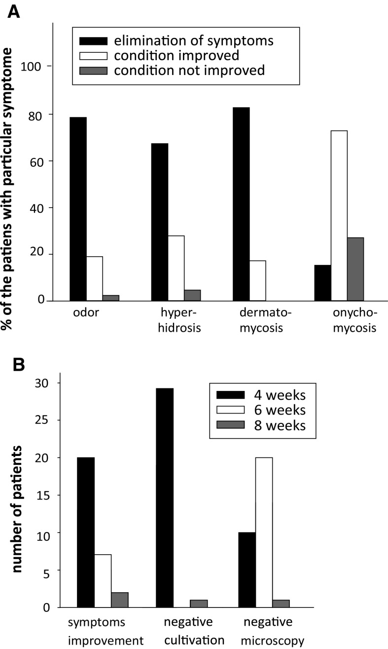
Pythium oligandrum (Oomycota) is known for its strong mycoparasitism against more than 50 fungal and oomycete species. However, the ability of this oomycete to suppress and kill the causal agents of dermatophytoses is yet to be studied. We provide a complex study of the interactions between P. oligandrum and dermatophytes representing all species dominating in the developed countries. We assessed its biocidal potential by performing growth tests, on both solid and liquid cultivation media and by conducting a pilot clinical study. In addition, we studied the molecular background of mycoparasitism using expression profiles of genes responsible for the attack on the side of P. oligandrum and the stress response on the side of Microsporum canis. We showed that dermatophytes are efficiently suppressed or killed by P. oligandrum in the artificial conditions of cultivations media between 48 and 72 h after first contact. Significant intra- and interspecies variability was noted. Of the 69 patients included in the acute regimen study, symptoms were completely eliminated in 79% of the patients suffering from foot odour, hyperhidrosis disappeared in 67% of cases, clinical signs of dermatomycoses could no longer be observed in 83% of patients, and 15% of persons were relieved of symptoms of onychomycosis. Our investigations provide clear evidence that the oomycete is able to recognize and kill dermatophytes using recognition mechanisms that resemble those described in oomycetes attacking fungi infecting plants, albeit with some notable differences.

Keywords: Aggressivity genes; Dermatophytes; Microsporum; Mycoparasitism; Pythium oligandrum; Trichophyton.


Conflict of interest statement

Conflict of interest

Martin Suchánek and Radim Klimeš are owners and stakeholders in the companies Biopreparáty and Bio Agens Research and Development manufacturing biological antifungal products based on Pythium oligandrum.

Research Involving Human Participants and/or Animals

For this type of study formal consent is not required.

Informed Consent

Informed consent was obtained from all individual participants included in the study.

Figures

Fig. 1
Fig. 1
Examples of the time course of direct interactions representing all five interaction patterns. Type I—exponential single phase of the ascending type. a Trichophyton rubrum CCF 4933. b Trichophyton benhamiae CCF 4918. Type II—exponential two-phase pattern of the ascending type. c Trichophyton erinacei CCF 4472. Type III—exponential single phase with an ascending and a descending phase. d. Nannizzia persicolor CCF 4542. e Epidermophyton floccosum PL 231. Type IV—two-phase pattern with an ascending and a descending phase. f. Nannizzia gypsea CCF 4626. The photograph was taken on days 4, 6, 8 and 10 of the experiment, ordered sequentially from left to right
Fig. 2
Fig. 2
Examples of the time course of the elimination of dermatophytes (gray line) by Pythium oligandrum (black line) on the MEA cultivation medium. a Epidermophyton floccosum. b Microsporum canis. c Nannizzia fulva d Nannizzia gypsea. e Nannizzia persicolor. f Trichophyton benhamiae. g Trichophyton erinacei. h Trichophyton interdigitale. i Trichophyton rubrum. j Trichophyton tonsurans. Error bars represent the standard deviation counted from all strains and Petri dishes of the particular dermatophyte species
Fig. 3
Fig. 3
Effects of the medium and strain on the competition between Pythium oligandrum and dermatophytes after 6 days of the experiment. Error bars represent the standard deviation counted from all strains and Petri dishes of the particular dermatophyte species
Fig. 4
Fig. 4
Gene expression profiles during the interaction of Pythium oligandrum with the dermatophyte Microsporum canis. An agar block with Pythium oligandrum was added on day 3 onto a Petri dish with a well-grown dermatophyte. Photodocumentation and gene expression profiling started from day 4 and proceeded until day 7. For Pythium oligandrum, we examined the expression of genes coding for cellulase (POCELL), endo-β-1,3-glucanase (POENDO) and the tyrosine-rich structural protein (POSTRU), whereas for Microsporum canis we followed the expression of genes for the LysM adhesion/masking protein (MCLYSM), metalloproteinase (MCMETA) and Ca-dependent kinase (MCCAMK). The results are shown at logarithmic scale; each bar shows the average value for three independent experiments
Fig. 5
Fig. 5
Gene expression profiles and viability during the interaction of Pythium oligandrum with the dermatophyte Microsporum canis in suspension. For Pythium oligandrum, we examined the expression of genes coding for cellulase (POCELL), endo-β-1,3-glucanase (POENDO) and the tyrosine-rich structural protein (POSTRU) whereas while for Microsporum canis, we followed the expression of genes for the LysM effector/masking protein (MCLYSM), metalloproteinase (MCMETA) and Ca-dependent kinase (MCCAMK). The results are shown at logarithmic scale; each bar shows the averaged value for three independent experiments
Fig. 6
Fig. 6
Efficacy of cosmetic products containing the oomycete Pythium oligandrum against symptoms of foot mycoses. a Elimination of individual symptoms in patients with acute patients. b Elimination of individual symptoms in patients with recurrent dermatophytoses

Similar articles

Cited by

References

    1. Weitzman I, Summerbell RC. The dermatophytes. Clin Microbiol Rev. 1995;8:240–259. - PMC - PubMed
    1. Achterman RR, White TC. A foot in the door for dermatophyte research. PLoS Pathog. 2012;8:e1002564. doi: 10.1371/journal.ppat.1002564. - DOI - PMC - PubMed
    1. Havlickova B, Czaika VA, Friedrich M. Epidemiological trends in skin mycoses worldwide. Mycoses. 2008;51:2–15. doi: 10.1111/j.1439-0507.2008.01606.x. - DOI - PubMed
    1. Drake LA, Dinehart SM, Farmer ER, Goltz RW, Graham GF, Hordinsky MK, et al. Guidelines of care for superficial mycotic infections of the skin: tinea corporis, tinea cruris, tinea faciei, tinea manuum, and tinea pedis. J Am Acad Dermatol. 1996;34:282–286. doi: 10.1016/S0190-9622(96)80135-6. - DOI - PubMed
    1. Achterman RR, White TC. Dermatophyte virulence factors: identifying and analyzing genes that may contribute to chronic or acute skin infections. Int J Microbiol. 2012;2012:358305. doi: 10.1155/2012/358305. - DOI - PMC - PubMed

LinkOut - more resources