. 2018 Aug;35(8):1419-1429.
doi: 10.1007/s10815-018-1231-4. Epub 2018 Jun 30.

Oviductal glycoprotein 1 (OVGP1) is expressed by endometrial epithelium that regulates receptivity and trophoblast adhesion

Affiliations

Oviductal glycoprotein 1 (OVGP1) is expressed by endometrial epithelium that regulates receptivity and trophoblast adhesion

Saniya Laheri et al. J Assist Reprod Genet. 2018 Aug.

Abstract

Purpose: To study the regulation and functions of oviductal glycoprotein 1 (OVGP1) in endometrial epithelial cells.

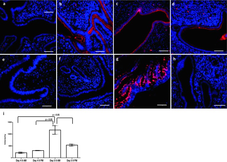
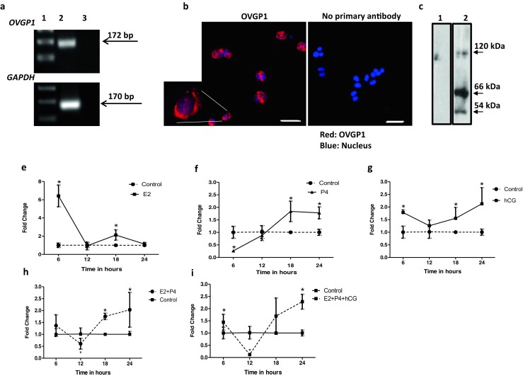
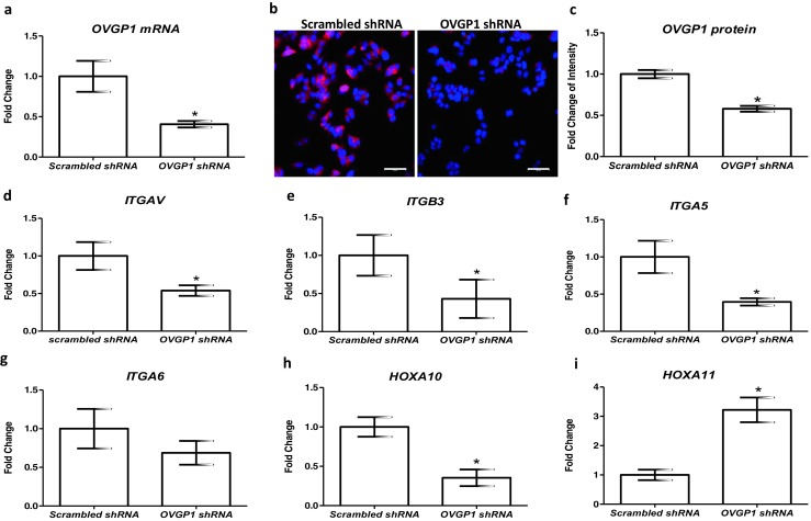
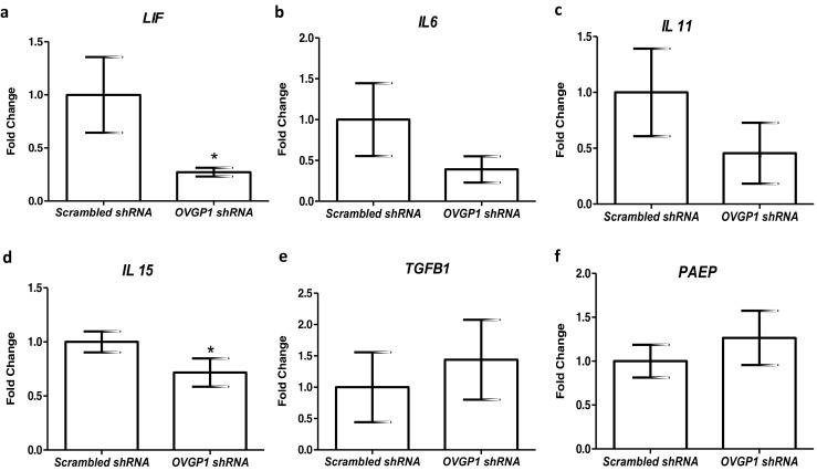
Methods: Expression of OVGP1 in mouse endometrium during pregnancy and in the endometrial epithelial cell line (Ishikawa) was studied by immunofluorescence, Western blotting, and RT-PCR. Regulation of OVGP1 in response to ovarian steroids and human chorionic gonadotropin (hCG) was studied by real-time RT-PCR. OVGP1 expression was knockdown in Ishikawa cells by shRNA, and expression of receptivity associated genes was studied by real-time RT-PCR. Adhesion of trophoblast cell line (JAr) was studied by in vitro adhesion assays.

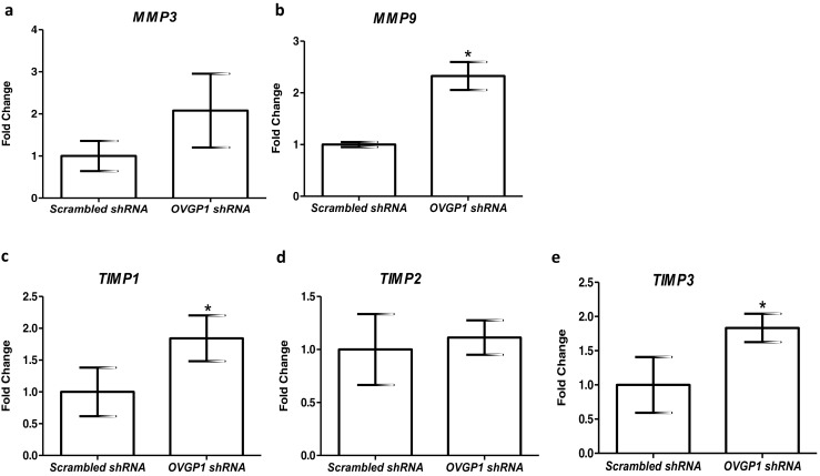
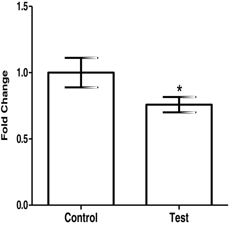
Results: OVGP1 was localized exclusively in the luminal epithelial cells of mouse endometrium at the time of embryo implantation. Along with estrogen and progesterone, hCG induced the expression of OVGP1 in Ishikawa cells. Knockdown of OVGP1 in Ishikawa cells reduced mRNA expression of ITGAV, ITGB3, ITGA5, HOXA10, LIF, and IL15; it increased the expression of HOXA11, MMP9, TIMP1, and TIMP3. Supernatants derived from OVGP1 knockdown Ishikawa cells reduced the adhesiveness of JAr cells in vitro. Expression of OVGP1 mRNA was found to be significantly lowered in the endometrium of women with recurrent implantation failure.

Conclusion: OVGP1 is specifically induced in the luminal epithelium at the time of embryo implantation where it regulates receptivity-related genes and aids in trophoblast adhesion.

Keywords: Endometrium; Glycoprotein; Implantation; Oviductal glycoprotein 1; Receptivity; Trophoblast.


Conflict of interest statement

The study was approved by the Institutional Animal Ethics Committee (IAEC) of the National Institute for Research in Reproductive (NIRRH).

The authors declare that they have no conflict of interests.

Figures

Fig. 1
Fig. 1
Expression of OVGP1 in mouse endometrium at time of embryo implantation. Paraffin sections were probed with OVGP1 antibody and detected with Alexa Fluor 568. a Day 4 morning. b Day 4 evening. c Day 5 morning. d Day 5 evening. e Estrus stage. f Diestrus stage. g Oviduct. h Negative control (without primary antibody). Red staining is for OVGP1, and blue is for the nuclei. Scale bar is 50 μm. i Intensity of OVGP1 staining during the course of embryo implantation. Y-axis is the intensity values for OVGP1 staining. Data is mean ± SD for the three biological replicates at each point. * indicates significant difference as compared to control (p < 0.05)
Fig. 2
Fig. 2
Effects of estrogen, progesterone, and human chorionic gonadotropin (hCG) on OVGP1 expression in human endometrial epithelial cells. a Expression of OVGP1 mRNA from Ishikawa cells. Lane 1 is 100 bp ladder, lane 2 cDNA from Ishikawa mRNA, and lane 3 no reverse transcriptase control. The bands for OVGP1 (172 bp) and GAPDH (170 bp) are shown by arrow marks. b Immunofluorescence for OVGP1 in Ishikawa cells. Red staining is for OVGP1, and blue is for the nuclei. Scale bar is 50 μm. A cell is digitally zoomed × 5 times and shown. c Western blotting for OVGP1 in protein isolated from Ishikawa cells. Lane 1 is negative control (incubated without primary antibody), and lane 2 is sample incubated with primary antibody. The approximate size of the bands is marked with an arrow. All the experiments were done at least three times. Ishikawa cells were treated with estrogen (E2), progesterone (P4), and hCG alone or in combinations (ei) for varying time points, and level of OVGP1 mRNA was measured by real-time PCR. Y-axis is fold change as compared to untreated control at each time point (control value taken as 1). X-axes are time points in hours post-treatment. Values are mean ± SD for six replicates. * indicates mean value significant different (p < 0.05) as compared to respective control
Fig. 3
Fig. 3
Effect of loss of OVGP1 on expression of receptivity markers in the endometrial epithelial cells. a mRNA levels of OVGP1 in scrambled and OVGP1 shRNA-transfected Ishikawa cells. Y-axis is fold change where values obtained from scrambled cells were taken as 1. b Immunofluorescence for OVGP1 in scrambled and knockdown Ishikawa cells. Red staining is for OVGP1, and blue is for the nuclei. Scale bar is 50 μm. c Intensity of OVGP1 staining in scrambled and OVGP1 shRNA-transfected Ishikawa cells. Y-axis is intensity values for OVGP1 staining. In both a and b, data is mean ± SD for the three independent replicates. * indicates significant difference as compared to control (p < 0.05). Effect of loss of OVGP1 on mRNA levels of ITGAV, ITGB3, ITGA5, ITGA6, HOXA10, and HOXA11 (di). Y-axis is fold change where values obtained from scrambled cells were taken as 1. Data is the mean ± SD for the three independent replicates. * indicates significant difference as compared to control (p < 0.05)
Fig. 4
Fig. 4
Effect of loss of OVGP1 on mRNA levels of LIF, IL6, IL11, IL15, TGFB1, and PAEP (af). Y-axis is fold change where values obtained from scrambled cells were taken as 1. Data is the mean ± SD for the three independent replicates. * indicates significant difference as compared to control (p < 0.05)
Fig. 5
Fig. 5
Effect of loss of OVGP1 on mRNA levels of MMP3, MMP9, TIMP1, TIMP2, and TIMP3 (ad). Y-axis is fold change where values obtained from scrambled cells were taken as 1. Data is the mean ± SD for three independent replicates. * indicates significant difference as compared to control (p < 0.05)
Fig. 6
Fig. 6
Effect of loss of OVGP1 in the endometrial epithelial cells on adhesion of trophoblast cells. Control is conditioned media from Ishikawa cells transfected with scrambled shRNA. Test is conditioned media from Ishikawa cells transfected with OVGP1 shRNA. Trophoblast cells (JAr cell line) were treated with the conditioned media for 24 h, trypsinized, and allowed to adhere for 6 h. The number of adherent cells was measured as described in the “Materials and methods” section. Values on Y indicate fold change where values obtained from control were taken as 1. Data are the mean ± SD of absorbance from two independent experiments done in triplicates. * indicates significant difference as compared to control (p < 0.05)

References

    1. Polanski LT, Baumgarten MN, Quenby S, Brosens J, Campbell BK, Raine-Fenning NJ. What exactly do we mean by ‘recurrent implantation failure’? A systematic review and opinion. Reprod BioMed Online. 2014;28(4):409–423. doi: 10.1016/j.rbmo.2013.12.006. - DOI - PubMed
    1. European IVF-monitoring Consortium Assisted reproductive technology in Europe, 2013: results generated from European registers by ESHRE. Hum Reprod. 2017;32(10):1957–1973. doi: 10.1093/humrep/dex264. - DOI - PubMed
    1. Cha J, Sun X, Dey SK. Mechanisms of implantation: strategies for successful pregnancy. Nat Med. 2012;18(12):1754–1767. doi: 10.1038/nm.3012. - DOI - PMC - PubMed
    1. Modi DN, Bhartiya P. Physiology of embryo-endometrial cross talk. Biomed Res J. 2015;2:83–104.
    1. Evans J, Salamonsen LA, Winship A, Menkhorst E, Nie G, Gargett CE, Dimitriadis E. Fertile ground: human endometrial programming and lessons in health and disease. Nat Rev Endocrinol. 2016;12(11):654–667. doi: 10.1038/nrendo.2016.116. - DOI - PubMed

LinkOut - more resources