

Polarized Secretion of Extracellular Vesicles by Mammary Epithelia
- PMID: 29968174
- PMCID: PMC6103817
- DOI: 10.1007/s10911-018-9402-6
Polarized Secretion of Extracellular Vesicles by Mammary Epithelia
Abstract
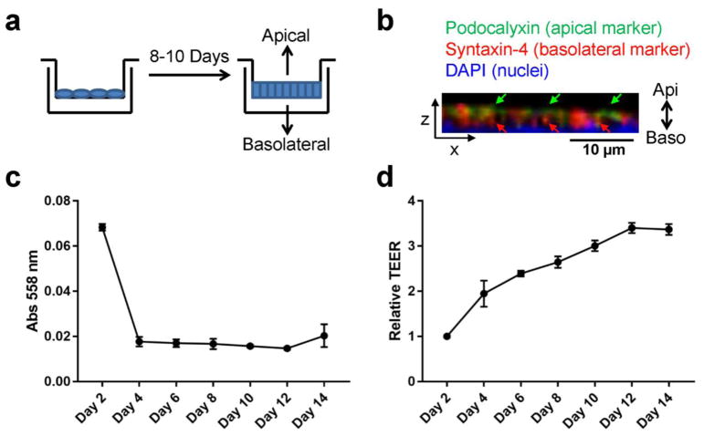
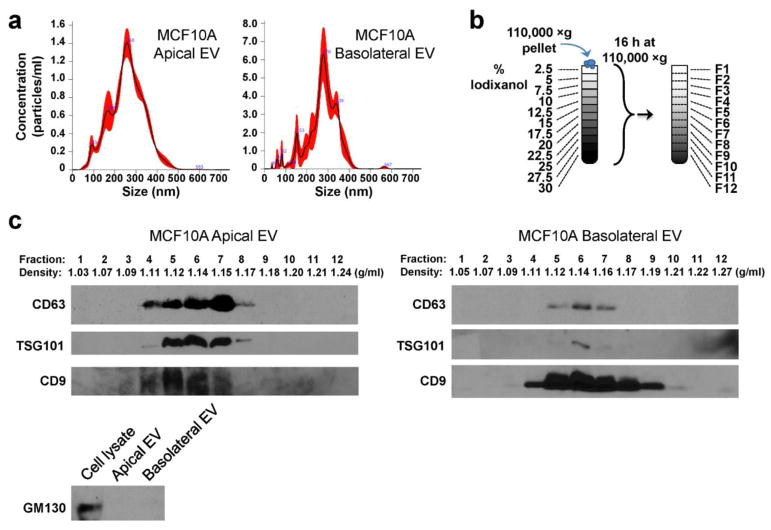
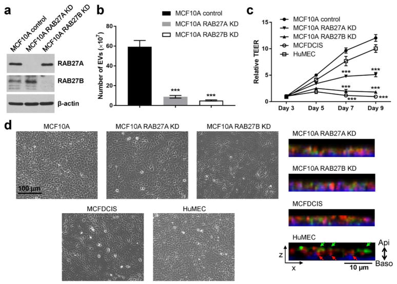
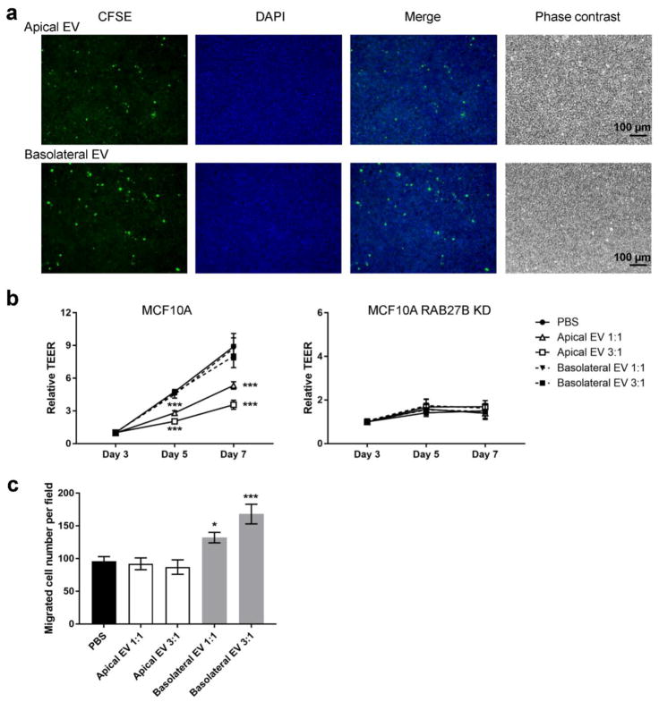
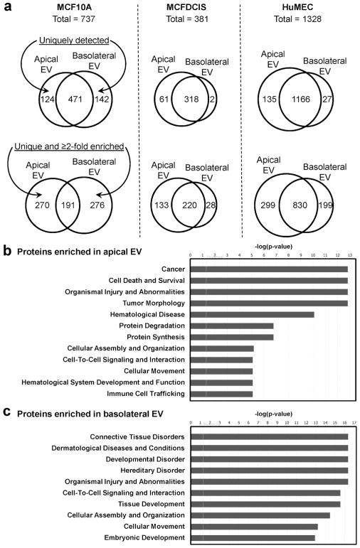
Extracellular vesicles (EVs) are secreted by many cell types and are increasingly investigated for their role in human diseases including cancer. Here we focus on the secretion and potential physiological function of non-pathological EVs secreted by polarized normal mammary epithelial cells. Using a transwell system to allow formation of epithelial polarity and EV collection from the apical versus basolateral compartments, we found that impaired secretion of EVs by knockdown of RAB27A or RAB27B suppressed the establishment of mammary epithelial polarity, and that addition of apical but not basolateral EVs suppressed epithelial polarity in a dose-dependent manner. This suggests that apical EV secretion contributes to epithelial polarity, and a possible mechanism is through removal of certain intracellular molecules. In contrast, basolateral but not apical EVs promoted migration of mammary epithelial cells in a motility assay. The protein contents of apical and basolateral EVs from MCF10A and primary human mammary epithelial cells were determined by mass spectrometry proteomic analysis, identifying apical-EV-enriched and basolateral-EV-enriched proteins that may contribute to different physiological functions. Most of these proteins differentially secreted by normal mammary epithelial cells through polarized EV release no longer showed polarized secretion in MCF10A-derived transformed epithelial cells. Our results suggest an essential role of EV secretion in normal mammary epithelial polarization and distinct protein contents and functions in apical versus basolateral EVs secreted by polarized mammary epithelia.
Keywords: Epithelial polarization; Extracellular vesicles; Mammary gland; Proteomics.
Conflict of interest statement
The authors declare no competing financial interests.
Figures
Similar articles
-
Polarized human cholangiocytes release distinct populations of apical and basolateral small extracellular vesicles.Mol Biol Cell. 2020 Oct 15;31(22):2463-2474. doi: 10.1091/mbc.E19-03-0133. Epub 2020 Aug 26. Mol Biol Cell. 2020. PMID: 32845745 Free PMC article.
-
Molecular and functional profiling of apical versus basolateral small extracellular vesicles derived from primary human proximal tubular epithelial cells under inflammatory conditions.J Extracell Vesicles. 2021 Feb;10(4):e12064. doi: 10.1002/jev2.12064. Epub 2021 Feb 16. J Extracell Vesicles. 2021. PMID: 33643548 Free PMC article.
-
Proteomic analysis of extracellular vesicles secreted by primary human epithelial endometrial cells reveals key proteins related to embryo implantation.Reprod Biol Endocrinol. 2022 Jan 3;20(1):3. doi: 10.1186/s12958-021-00879-x. Reprod Biol Endocrinol. 2022. PMID: 34980157 Free PMC article.
-
Cell polarity in motion: redefining mammary tissue organization through EMT and cell polarity transitions.J Mammary Gland Biol Neoplasia. 2010 Jun;15(2):149-68. doi: 10.1007/s10911-010-9180-2. Epub 2010 May 12. J Mammary Gland Biol Neoplasia. 2010. PMID: 20461450 Review.
-
Tissue polarity-dependent control of mammary epithelial homeostasis and cancer development: an epigenetic perspective.J Mammary Gland Biol Neoplasia. 2010 Mar;15(1):49-63. doi: 10.1007/s10911-010-9168-y. Epub 2010 Jan 27. J Mammary Gland Biol Neoplasia. 2010. PMID: 20101444 Free PMC article. Review.
Cited by
-
Assessing extracellular vesicles from bovine mammary gland epithelial cells cultured in FBS-free medium.Extracell Vesicles Circ Nucl Acids. 2021 Dec 31;2(4):252-267. doi: 10.20517/evcna.2021.18. eCollection 2021. Extracell Vesicles Circ Nucl Acids. 2021. PMID: 39697662 Free PMC article.
-
Challenges for Studying and Isolating Extracellular Vesicles from Cell-Conditioned Media.Methods Mol Biol. 2023;2666:299-315. doi: 10.1007/978-1-0716-3191-1_21. Methods Mol Biol. 2023. PMID: 37166673
-
Extracellular vesicles in dairy cattle: research progress and prospects for practical applications.J Anim Sci Biotechnol. 2025 Aug 4;16(1):110. doi: 10.1186/s40104-025-01242-5. J Anim Sci Biotechnol. 2025. PMID: 40754626 Free PMC article. Review.
-
Polarized human cholangiocytes release distinct populations of apical and basolateral small extracellular vesicles.Mol Biol Cell. 2020 Oct 15;31(22):2463-2474. doi: 10.1091/mbc.E19-03-0133. Epub 2020 Aug 26. Mol Biol Cell. 2020. PMID: 32845745 Free PMC article.
-
Mesenchymal Stem Cell-Derived Extracellular Vesicles: Regenerative Potential and Challenges.Biology (Basel). 2021 Feb 25;10(3):172. doi: 10.3390/biology10030172. Biology (Basel). 2021. PMID: 33668707 Free PMC article. Review.
References
Publication types
MeSH terms
Substances
Grants and funding
LinkOut - more resources
Full Text Sources
Other Literature Sources
