. 2018 Oct;246(2):180-190.
doi: 10.1002/path.5130. Epub 2018 Aug 22.

Activation of sterol regulatory element-binding protein 1 (SREBP1)-mediated lipogenesis by the Epstein-Barr virus-encoded latent membrane protein 1 (LMP1) promotes cell proliferation and progression of nasopharyngeal carcinoma

Affiliations

Activation of sterol regulatory element-binding protein 1 (SREBP1)-mediated lipogenesis by the Epstein-Barr virus-encoded latent membrane protein 1 (LMP1) promotes cell proliferation and progression of nasopharyngeal carcinoma

Angela Kwok-Fung Lo et al. J Pathol. 2018 Oct.

Abstract

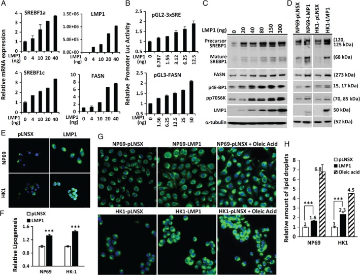
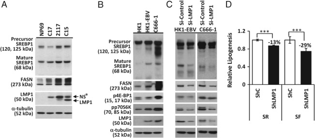
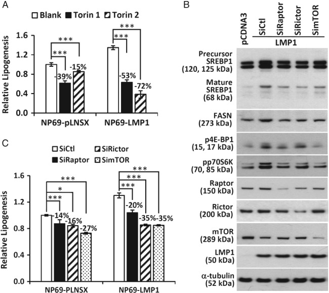
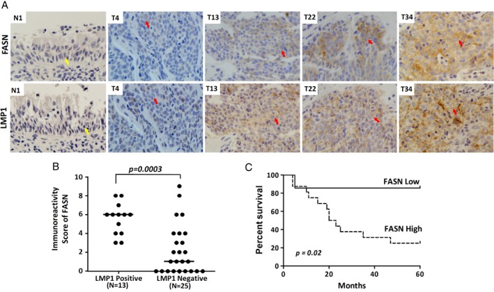
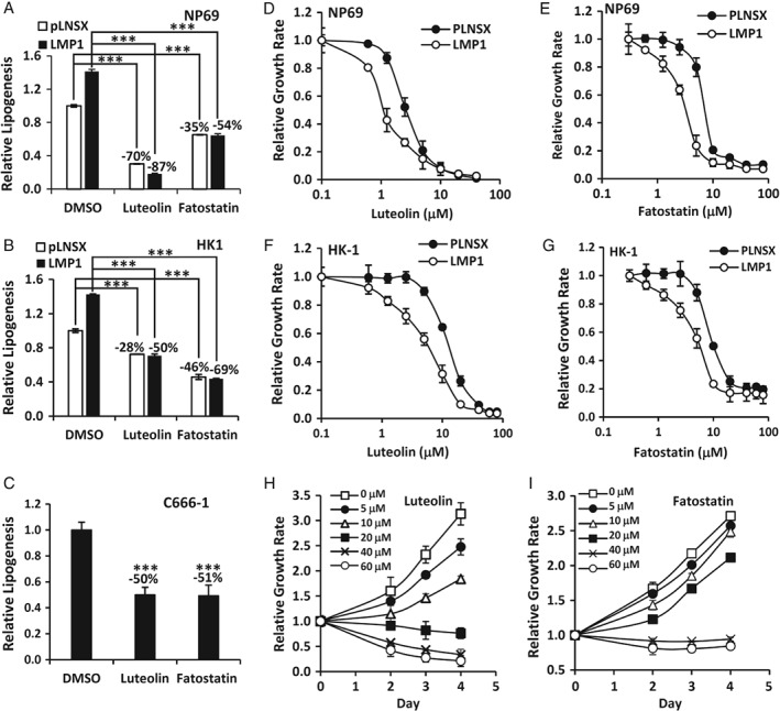
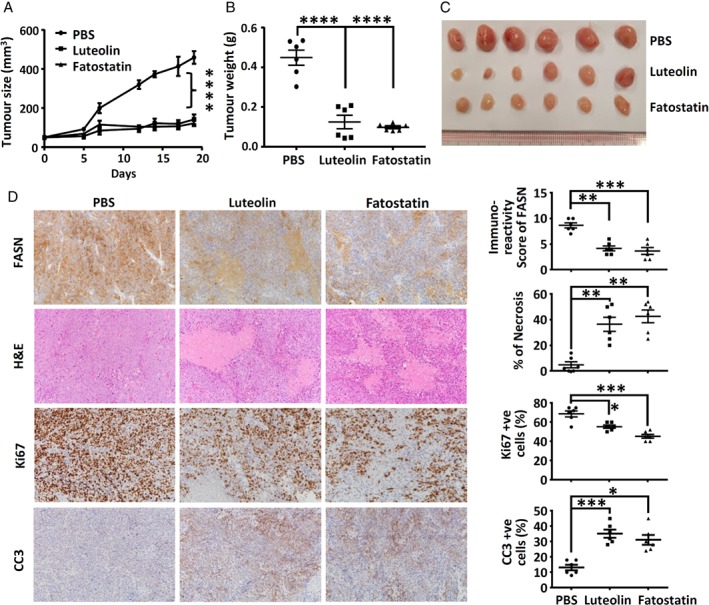
Nasopharyngeal carcinoma (NPC) is closely associated with Epstein-Barr virus (EBV) infection. The EBV-encoded latent membrane protein 1 (LMP1), which is commonly expressed in NPC, engages multiple signaling pathways that promote cell growth, transformation, and metabolic reprogramming. Here, we report a novel function of LMP1 in promoting de novo lipogenesis. LMP1 increases the expression, maturation and activation of sterol regulatory element-binding protein 1 (SREBP1), a master regulator of lipogenesis, and its downstream target fatty acid synthase (FASN). LMP1 also induces de novo lipid synthesis and lipid droplet formation. In contrast, small interfering RNA (siRNA) knockdown of LMP1 in EBV-infected epithelial cells diminished SREBP1 activation and lipid biosynthesis. Furthermore, inhibition of the mammalian target of rapamycin (mTOR) pathway, through the use of either mTOR inhibitors or siRNAs, significantly reduced LMP1-mediated SREBP1 activity and lipogenesis, indicating that LMP1 activation of the mTOR pathway is required for SREBP1-mediated lipogenesis. In primary NPC tumors, FASN overexpression is common, with high levels correlating significantly with LMP1 expression. Moreover, elevated FASN expression was associated with aggressive disease and poor survival in NPC patients. Luteolin and fatostatin, two inhibitors of lipogenesis, suppressed lipogenesis and proliferation of nasopharyngeal epithelial cells, effects that were more profound in cells expressing LMP1. Luteolin and fatostatin also dramatically inhibited NPC tumor growth in vitro and in vivo. Our findings demonstrate that LMP1 activation of SREBP1-mediated lipogenesis promotes tumor cell growth and is involved in EBV-driven NPC pathogenesis. Our results also reveal the therapeutic potential of utilizing lipogenesis inhibitors in the treatment of locally advanced or metastatic NPC. © 2018 The Authors. The Journal of Pathology published by John Wiley & Sons Ltd on behalf of Pathological Society of Great Britain and Ireland.

Keywords: Epstein-Barr virus; LMP1; SREBP1; lipogenesis; nasopharyngeal carcinoma.


Figures

Figure 1
Figure 1
Induction of SREBP1 expression and activity by LMP1. (A) NP69 cells transfected with increasing amounts of an LMP1 expression vector as indicated were subjected to RT‐qPCR analysis for SREBF1a, SREBF1c, LMP1, and FASN. The mRNA expression of the target gene of interest was normalized to expression of TBP. Relative mRNA levels were calculated with the sample without the LMP1 expression vector (set at 1). (B) HEK‐293 cells were transfected with various amounts of an LMP1 expression vector together with luciferase promoter vector: pGL2‐3xSRE or pGL3‐FASN. After serum deprivation for 12 h, cells were harvested for reporter activity analysis. The firefly luciferase activity was normalized to Renilla luciferase activity, and plotted relative to the sample without the LMP1 expression vector (set at 1). (C and D) NP69 cells transfected with increasing amounts of an LMP1 expression vector as indicated (C) or NP69 and HK1 cells stably expressing LMP1 (D) were incubated in serum‐free medium for 12 h, prior to immunoblotting analysis. (E) Immunofluorescence staining of FASN. (F) [14C]Acetate incorporation assay for the measurement of de novo lipid synthesis. (G) Nile Red fluorescence staining for lipid droplets and counterstaining with Hoechst 33342 to stain cell nuclei. As a positive control for lipid droplet staining (right panel), pLNSX control cells were incubated in medium supplemented with oleic acid for 12 h prior to staining. Nile Red fluorescence (excitation, 385 nm; emission, 535 nm) and Hoechst 33342 fluorescence (excitation, 355 nm; emission, 460 nm). (H) The relative amount of lipid droplet formation was calculated by normalizing the Hoechst 33342 fluorescence to the Nile Red signal in each well, and compared with the pLNSX vector control sample (set at 1). Data represent the means of eight determinations. Mean and standard deviation. ***p < 0.001.
Figure 2
Figure 2
LMP1 induction of SREBP1‐mediated lipogenesis in EBV‐infected NPC cells. (A) NPC xenografts (C17, 2117, and C15) together with cells of the immortalized nasopharyngeal epithelial cell line, NP69, which were incubated in serum‐free medium for 16 h prior to harvesting, were subjected to western blotting analysis for the indicated proteins. (B) The EBV‐infected NPC cell lines HK1‐EBV and C666‐1, together with EBV‐negative HK‐1 cells, were incubated in serum‐free medium for 12 h prior to western blotting analysis. (C) HK1‐EBV and C666‐1 cells transfected with negative siRNA control (si‐Control) or siRNA targeting LMP1 (si‐LMP1) were subjected to western blotting analysis. (D) C666‐1 cells expressing pSuper.retro‐shRNA scrambled control (ShC) or shRNA LMP1 (ShLMP1) were incubated in serum‐rich (SR) or serum‐free (SF) medium containing 2.5 μCi/ml [1‐14C]acetate for 10 h, and then subjected to measurement of lipid synthesis. NS*, non‐specific band. Mean and standard deviation. ***p < 0.001.
Figure 3
Figure 3
LMP1 activation of the mTOR signaling pathway is required for SREBP1‐mediated lipogenesis. (A) NP69‐pLNSX and NP69‐LMP1 cells treated with vehicle alone (Blank), Torin 1 (0.3 μm) or Torin 2 (0.1 μm). (B) NP69 cells transfected with either control pCDNA3 vector or pCDNA3‐LMP1 expression vector, together with a control scrambled siRNA (siCtl), or siRNA specific for Raptor (siRaptor), Rictor (siRictor), or mTOR (simTOR), were subjected to western blotting analysis for the indicated proteins. (C) NP69‐pLNSX and NP69‐LMP1 cells transfected with the indicated siRNAs for 24 h were subjected to lipid synthesis measurements. The asterisks indicate a significant difference (*p < 0.05, **p < 0.01, ***p < 0.001).
Figure 4
Figure 4
Elevation of FASN expression correlates with LMP1 expression in NPC tumors and is associated with poor prognosis in NPC patients. (A) Immunohistochemical staining of FASN and LMP1 in primary NPC and normal nasopharyngeal epithelium (NE) specimens. An absence of FASN and LMP1 expression was observed in NE (N1). In NPC tumor T4, which is LMP1‐negative, low levels of FASN (immunoreactivity score of 1) were observed. In NPC tumors T13, T22, and T34, which are LMP1‐positive, moderate (immunoreactivity score of 4) and high (immunoreactivity scores of 6 and 8) levels of FASN were observed. Yellow arrow: normal nasopharyngeal epithelium. Red arrow: NPC tumor cells. (B) Dot plot showing the immunoreactivity scores of FASN staining within the group of tumors with and without LMP1 expression (N = 38). All LMP1‐positive NPC tumors (13/13) showed strong FASN expression (immunoreactivity score of ≥3). The median value of each group is shown by the horizontal line. The p‐value between two groups is shown. (C) Kaplan–Meier survival curves for NPC patients with available follow‐up information. High FASN expression: immunoreactivity score of ≥3, n = 16. Low FASN expression: immunoreactivity score of <3, n = 7.
Figure 5
Figure 5
LMP1 induction of lipogenesis contributes to cell proliferation. (A–C) NP69‐pLNSX and NP69‐LMP1 cells (A), HK1‐pLNSX and HK1‐LMP1 cells (B) and C6661‐1 EBV‐positive NPC cells (C) cultured with 1% serum were treated with dimethylsulfoxide (DMSO) (vehicle), luteolin (20 μm) or fatostatin (15 μm) for 16 h prior to lipogenesis measurements. Relative lipogenesis was calculated with vehicle‐treated cells as the reference control (set at 1). (D–I) NP69‐pLNSX and NP69‐LMP1 cells (D and E), HK1‐pLNSX and HK1‐LMP1 cells (F and G) and C666‐1 cells (H and I) were treated with increasing doses of inhibitors, as indicated, for 3 days prior to cell growth analysis. Relative cell growth was calculated with vehicle‐treated cells as the reference control (set at 1). ***p < 0.001.
Figure 6
Figure 6
Suppression of NPC tumor growth by inhibitors of lipogenesis. Mice bearing C666‐1 xenografts were treated with PBS (control), luteolin (20 mg/kg) or fatostatin (15 mg/kg) every 2–3 days for 3 weeks. (A) Tumor size was measured every 2–3 days for 19 days. (B and C) At the endpoint, mice were killed, and tumors were (B) weighed and (C) photographed. (D, left panel) The harvested xenografts were embedded for hematoxylin and eosin (H&E) staining and immunohistochemical staining analysis. (D, right panel) The percentage of necrosis, and positive staining for FASN, Ki67, and CC3, were measured, and are shown as dot‐plots. Lines and bars are mean and standard error of the mean of each experimental group. *p < 0.05, **p < 0.01, ***p < 0.001, ****p < 0.0001.

Similar articles

Cited by

References

    1. Tsao SW, Tsang CM, To KF , et al The role of Epstein–Barr virus in epithelial malignancies. J Pathol 2015; 235 : 323–333. - PMC - PubMed
    1. Young LS, Dawson CW. Epstein–Barr virus and nasopharyngeal carcinoma. Chin J Cancer 2014; 33 : 581–590. - PMC - PubMed
    1. Dawson CW, Port RJ, Young LS. The role of the EBV‐encoded latent membrane proteins LMP1 and LMP2 in the pathogenesis of nasopharyngeal carcinoma (NPC). Semin Cancer Biol 2012; 22 : 144–153. - PubMed
    1. Lo AK, Lo KW, Ko CW, et al Inhibition of the LKB1–AMPK pathway by the Epstein–Barr virus‐encoded LMP1 promotes proliferation and transformation of human nasopharyngeal epithelial cells. J Pathol 2013; 230 : 336–346. - PubMed
    1. Lo AK, Dawson CW, Young LS, et al Activation of the FGFR1 signalling pathway by the Epstein–Barr virus‐encoded LMP1 promotes aerobic glycolysis and transformation of human nasopharyngeal epithelial cells. J Pathol 2015; 237 : 238–248. - PubMed

Publication types

MeSH terms

Substances