

Flow-metabolism uncoupling in patients with asymptomatic unilateral carotid artery stenosis assessed by multi-modal magnetic resonance imaging
- PMID: 29968499
- PMCID: PMC6827123
- DOI: 10.1177/0271678X18783369
Flow-metabolism uncoupling in patients with asymptomatic unilateral carotid artery stenosis assessed by multi-modal magnetic resonance imaging
Abstract
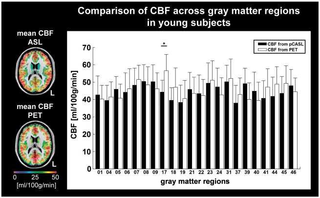
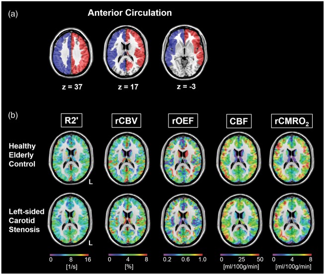
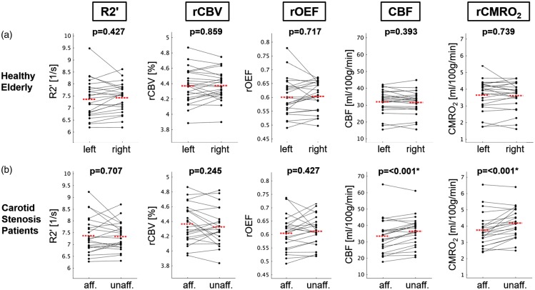
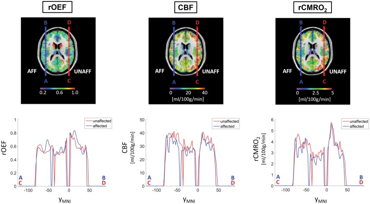
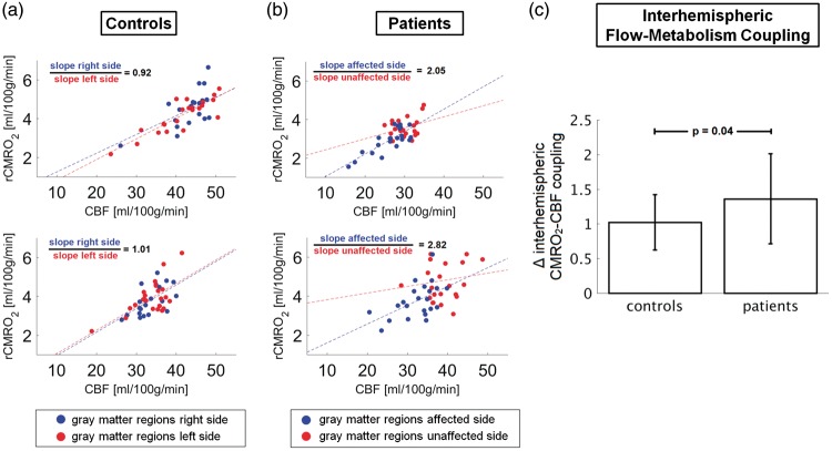
Oxygen extraction (OEF), oxidative metabolism (CMRO2), and blood flow (CBF) in the brain, as well as the coupling between CMRO2 and CBF due to cerebral autoregulation are fundamental to brain's health. We used a clinically feasible MRI protocol to assess impairments of these parameters in the perfusion territories of stenosed carotid arteries. Twenty-nine patients with unilateral high-grade carotid stenosis and thirty age-matched healthy controls underwent multi-modal MRI scans. Pseudo-continuous arterial spin labeling (pCASL) yielded absolute CBF, whereas multi-parametric quantitative blood oxygenation level dependent (mqBOLD) modeling allowed imaging of relative OEF and CMRO2. Both CBF and CMRO2 were significantly reduced in the stenosed territory compared to the contralateral side, while OEF was evenly distributed across both hemispheres similarly in patients and controls. The CMRO2-CBF coupling was significantly different between both hemispheres in patients, i.e. significant interhemispheric flow-metabolism uncoupling was observed in patients compared to controls. Given that CBF and CMRO2 are intimately linked to brain function in health and disease, the proposed easily applicable MRI protocol of pCASL and mqBOLD imaging might serve as a valuable tool for early diagnosis of potentially harmful cerebral hemodynamic and metabolic states with the final aim to select clinically asymptomatic patients who would benefit from carotid revascularization therapy.
Keywords: Carotid stenosis; arterial spin labeling; cerebral blood flow; cerebral metabolic rate of oxygen consumption; multi-parametric quantitative blood oxygenation level dependent imaging; perfusion.
Figures
Similar articles
-
Bilateral Cerebral Hypoperfusion in Asymptomatic Unilateral Carotid Artery Stenosis: An Arterial Spin Labeling MRI Study.Medicina (Kaunas). 2025 Apr 22;61(5):771. doi: 10.3390/medicina61050771. Medicina (Kaunas). 2025. PMID: 40428729 Free PMC article.
-
Trajectories and sex differences of brain structure, oxygenation and perfusion functions in normal aging.Neuroimage. 2024 Nov 15;302:120903. doi: 10.1016/j.neuroimage.2024.120903. Epub 2024 Oct 24. Neuroimage. 2024. PMID: 39461605
-
Structural MRI of carotid artery atherosclerotic lesion burden and characterization of hemispheric cerebral blood flow before and after carotid endarterectomy.NMR Biomed. 2006 Apr;19(2):198-208. doi: 10.1002/nbm.1017. NMR Biomed. 2006. PMID: 16475206 Clinical Trial.
-
Blood oxygenation level-dependent (BOLD)-based techniques for the quantification of brain hemodynamic and metabolic properties - theoretical models and experimental approaches.NMR Biomed. 2013 Aug;26(8):963-86. doi: 10.1002/nbm.2839. Epub 2012 Aug 28. NMR Biomed. 2013. PMID: 22927123 Free PMC article. Review.
-
Cerebral oxygen extraction fraction MRI: Techniques and applications.Magn Reson Med. 2022 Aug;88(2):575-600. doi: 10.1002/mrm.29272. Epub 2022 May 5. Magn Reson Med. 2022. PMID: 35510696 Free PMC article. Review.
Cited by
-
Spatiotemporal features of neurovascular (un)coupling with stimulus-induced activity and hypercapnia challenge in cerebral cortex and olfactory bulb.J Cereb Blood Flow Metab. 2023 Nov;43(11):1891-1904. doi: 10.1177/0271678X231183887. Epub 2023 Jun 21. J Cereb Blood Flow Metab. 2023. PMID: 37340791 Free PMC article.
-
Imaging effective oxygen diffusivity in the human brain with multiparametric magnetic resonance imaging.J Cereb Blood Flow Metab. 2022 Feb;42(2):349-363. doi: 10.1177/0271678X211048412. Epub 2021 Sep 30. J Cereb Blood Flow Metab. 2022. PMID: 34590895 Free PMC article. Clinical Trial.
-
Mapping oxidative metabolism in the human brain with calibrated fMRI in health and disease.J Cereb Blood Flow Metab. 2022 Jul;42(7):1139-1162. doi: 10.1177/0271678X221077338. Epub 2022 Mar 16. J Cereb Blood Flow Metab. 2022. PMID: 35296177 Free PMC article.
-
Assessment of cerebral autoregulatory function and inter-hemispheric blood flow in older adults with internal carotid artery stenosis using transcranial Doppler sonography-based measurement of transient hyperemic response after carotid artery compression.Geroscience. 2023 Dec;45(6):3333-3357. doi: 10.1007/s11357-023-00896-1. Epub 2023 Aug 21. Geroscience. 2023. PMID: 37599343 Free PMC article.
-
Neuroimaging anomalies in asymptomatic middle cerebral artery steno-occlusive disease with normal-appearing white matter.Front Neurol. 2023 Aug 24;14:1206786. doi: 10.3389/fneur.2023.1206786. eCollection 2023. Front Neurol. 2023. PMID: 37693758 Free PMC article.
References
-
- Derdeyn CP, Videen TO, Yundt KD, et al. Variability of cerebral blood volume and oxygen extraction: stages of cerebral haemodynamic impairment revisited. Brain 2002; 125(Pt 3): 595–607. - PubMed
-
- Derdeyn CP, Videen TO, Grubb RL, Jr, et al. Comparison of PET oxygen extraction fraction methods for the prediction of stroke risk. J Nucl Med 2001; 42: 1195–1197. - PubMed
-
- Yamauchi H, Fukuyama H, Nagahama Y, et al. Significance of increased oxygen extraction fraction in five-year prognosis of major cerebral arterial occlusive diseases. J Nucl Med 1999; 40: 1992–1998. - PubMed
-
- ten Dam VH, van den Heuvel DM, de Craen AJ, et al. Decline in total cerebral blood flow is linked with increase in periventricular but not deep white matter hyperintensities. Radiology 2007; 243: 198–203. - PubMed
Publication types
MeSH terms
Substances
Grants and funding
LinkOut - more resources
Full Text Sources
Other Literature Sources
Medical
