. 2018 Jul 3;8(1):10048.
doi: 10.1038/s41598-018-28443-5.

Cold atmospheric plasma causes a calcium influx in melanoma cells triggering CAP-induced senescence

Affiliations

Cold atmospheric plasma causes a calcium influx in melanoma cells triggering CAP-induced senescence

Christin Schneider et al. Sci Rep. .

Abstract

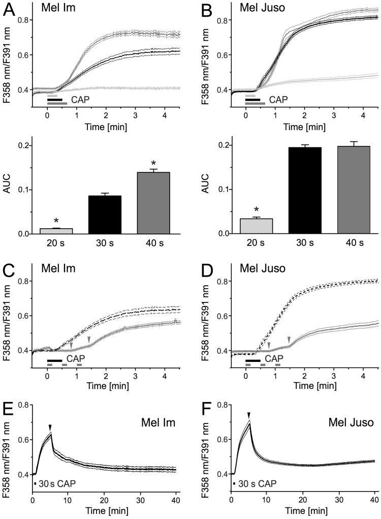
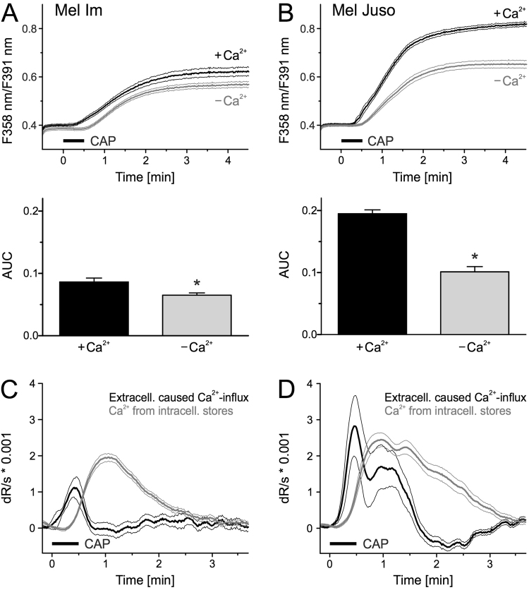
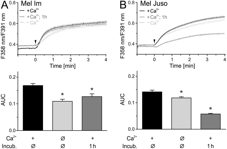
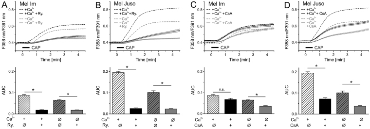
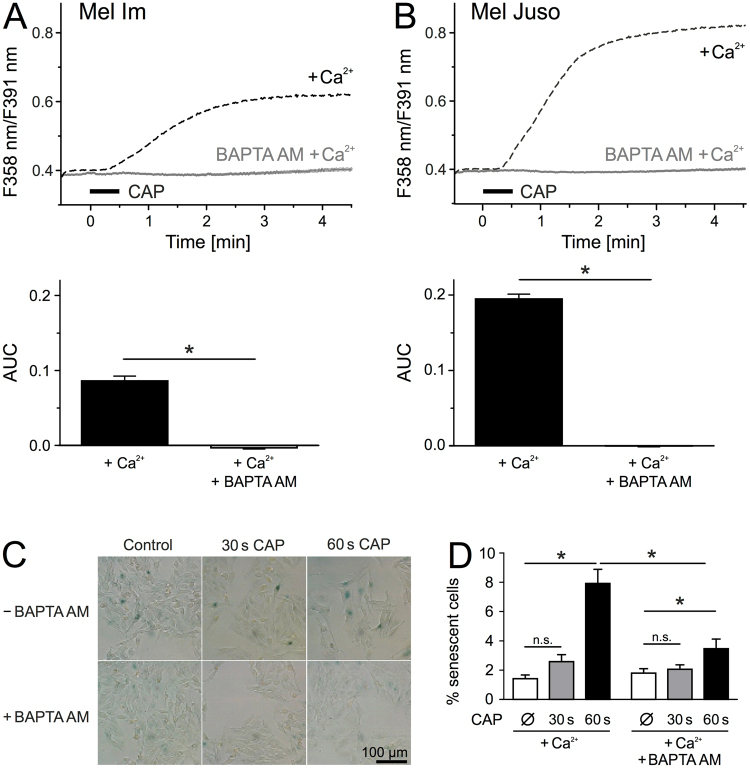
Cold atmospheric plasma (CAP) is a promising approach in anti-cancer therapy, eliminating cancer cells with high selectivity. However, the molecular mechanisms of CAP action are poorly understood. In this study, we investigated CAP effects on calcium homeostasis in melanoma cells. We observed increased cytoplasmic calcium after CAP treatment, which also occurred in the absence of extracellular calcium, indicating the majority of the calcium increase originates from intracellular stores. Application of previously CAP-exposed extracellular solutions also induced cytoplasmic calcium elevations. A substantial fraction of this effect remained when the application was delayed for one hour, indicating the chemical stability of the activating agent(s). Addition of ryanodine and cyclosporin A indicate the involvement of the endoplasmatic reticulum and the mitochondria. Inhibition of the cytoplasmic calcium elevation by the intracellular chelator BAPTA blocked CAP-induced senescence. This finding helps to understand the molecular influence and the mode of action of CAP on tumor cells.


Conflict of interest statement

The authors declare no competing interests.

Figures

Figure 1
Figure 1
CAP treatment leads to a delayed Ca2+ influx. Cytoplasmic Ca2+ time courses were measured using fura-2 AM. CAP exposure for 20 s, 30 s and 40 s elevated cytoplasmic Ca2+ in Mel Im (A, n = 500–660) and Mel Juso (B, n = 280–503). The area under the curve (AUC) of the first 90 s of this response was quantified in the bar charts. (C) Mel Im (n = 299) and (D) Mel Juso (n = 261) show the response pattern more clearly when the 30 s exposure (as in A,B for reference, dotted line) is fractionated into 3 times with 10 s CAP as indicated by the grey bars. The grey arrows mark the delay of the Ca2+ response. (E,F) Long-term measurement of cytoplasmic Ca2+ after treatment of Mel Im ((E), n = 275) and Mel Juso ((F), n = 353) with 30 s CAP. PbECS (1 ml) was added 5 minutes after the start of recording, represented as black arrows. Data are shown as mean and 99% confidence interval. N indicates the investigated number of cells.
Figure 2
Figure 2
CAP induces Ca2+ primarily from intracellular sources. (A,B) Cytoplasmic Ca2+ levels stimulated by 30 s CAP exposure in the presence or absence of extracellular Ca2+. Mel Im ((A), n = 660 and 464) and Mel Juso. ((B), n = 492 and 397) were washed for 5 min in pbECS before CAP in a solution with or without Ca2+. (C,D) Smoothed first derivative (dR/dt) of experiment without extracellular Ca2+ (grey trace). The effect due to the presence of extracellular Ca2+ is calculated from the difference of experiment with and without extracellular Ca2+ (black trace). Data are shown as mean and 99% confidence interval.
Figure 3
Figure 3
Addition of CAP-pretreated solution onto cells leads to an immediate Ca2+ influx. Measurement of cytoplasmic Ca2+ using fura-2 AM. CAP exposed pbECS (100 µl) with Ca2+ was applied (black traces) onto Mel Im ((A), n = 485) and Mel Juso ((B), n = 321) 1 min after start of recording (arrowhead). The experiment was repeated with an interval of 1 h between CAP-exposure and application of the solutions (dark grey traces) and in the absence of extracellular Ca2+ (light grey trace) ((A), n = 274–411; (B), n = 366–579). Data are shown as mean and 99% confidence interval.
Figure 4
Figure 4
The endoplasmic reticulum and the mitochondria are involved in CAP-induced Ca2+ response. Cytoplasmic Ca2+ responses induced by 30 s CAP exposure in Mel Im and Mel Juso were reduced by ryanodine (Ry., 30 µM; ((A,B), n = 324–587) or cyclosporin A (CsA, 0.5 µM; ((C,D), n = 349–516). Experiments were performed in presence or absence of extracellular Ca2+. Data are shown as mean and 99% confidence interval.
Figure 5
Figure 5
CAP-induced senescence and Ca2+ influx are linked to each other. (A,B) Mel Im (A, n = 389) and Mel Juso (B, n = 660) were simultaneously loaded with the Ca2+ chelator BAPTA AM (10 µM) and fura-2 AM (3 µM) and cytoplasmic Ca2+ was measured during and after 30 s CAP exposure. (C,D) Senescence-associated ß-galactosidase staining of Mel Juso (n = 20, each from four independent runs) 48 h after CAP exposure for 30 s or 60 s and with or without BAPTA AM (10 µM) pretreatment. (C) Representative pictures were taken with a 20 × magnification in bright field illumination. Senescent cells showed a blue staining due to senescence-associated ß-galactosidase activity. Traces are mean and 99% confidence interval (A,B) and bars are mean ± SEM (D).

Similar articles

Cited by

References

    1. Weltmann K-D, von Woedtke T. Plasma medicine—current state of research and medical application. Plasma Phys. Control. Fusion. 2017;59:14031. doi: 10.1088/0741-3335/59/1/014031. - DOI
    1. Hoffmann, C., Berganza, C. & Zhang, J. Cold Atmospheric Plasma: Methods of production and application in dentistry and oncology. Med. Gas Res. 3, (2013). - PMC - PubMed
    1. Heinlin J, et al. Plasma applications in medicine with a special focus on dermatology. J. Eur. Acad. Dermatology Venereol. 2011;25:1–11. doi: 10.1111/j.1468-3083.2010.03702.x. - DOI - PubMed
    1. Heinlin J, et al. Plasma medicine: possible applications in dermatology. JDDG - J. Ger. Soc. Dermatology. 2010;8:968–977. - PubMed
    1. Fridman G, et al. Applied plasma medicine. Plasma Process. Polym. 2008;5:503–533. doi: 10.1002/ppap.200700154. - DOI

Publication types