Case Reports
doi: 10.15537/smj.2018.7.22265.
Untreated submandibular megalith for over 60 years
Affiliations
- PMID: 29968898
- PMCID: PMC6146257
- DOI: 10.15537/smj.2018.7.22265
Item in Clipboard
Case Reports
Untreated submandibular megalith for over 60 years
Saudi Med J.
2018 Jul.
Abstract
Intra-parenchymal sialolithiasis and subsequent fibrosis of the submandibular salivary glands is a rare disorder. The resulting swelling, pain, and infection derives affected patients to seek treatment. We present the case of an 85-years-old Saudi male patient who suffered from repeated swelling and infection in the left submandibular region which was misdiagnosed and treated for over 60 years as dental infection, infected skin sebaceous gland or lipoma. The presented case represents the largest intra-glandular submandibular stone with the longest duration ever reported in the medical literature.
Figures

Clinical extra-oral view showing submandibular swelling of the left side of the neck.

Ortho-pantomo-graphic (OPG) x-ray view at the time of presentation showing a calcified mass in the left submandibular region (arrow).

Ortho-pantomo-graphic (OPG) x-ray view 12 years earlier to the time of presentation showing a calcified mass in the left submandibular region (white arrow).

CT coronal view showing a salivary stone within the left submandibular salivary gland.
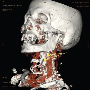
CT 3D view showing left submandibular salivary stone (arrows) with smooth surface caudal extension closely related to the hyoid bone.

CT 3D view showing the left submandibular salivary stone (arrow) and its relation to adjacent structures without any bony invasion.

Patient’s timeline.
References
-
- Abdullah O, AlQudehy Z. Giant submandibular sialolith: A case report and literature review. Indian Journal of Otology. 2016;22:126–128.
-
- Kim JP, Park JJ, Son HY, Woo SW. An Unusual Case of Bilateral Submandibular Sialolithiasis. Journal of Medical Cases. 2012;3:106–109.
-
- Kamtane S, Ghodke M. Sialolithiasis: An Unusually Large Asymptomatic Submandibular Salivary Stone. Serbian Dental Journal. 2013;60(1)
-
- Zenk J, Koch M, Klintworth N, König B, Konz K, Gillespie MB, et al. Sialendoscopy in the diagnosis and treatment of sialolithiasis: a study on more than 1000 patients. Otolaryngol Head Neck Surg. 2012;147:858–863. - PubMed
Publication types
MeSH terms
LinkOut - more resources
Full Text Sources
Other Literature Sources