

Crystallization and X-ray analysis of all of the players in the autoregulation of the ataRT toxin-antitoxin system
- PMID: 29969102
- PMCID: PMC6038448
- DOI: 10.1107/S2053230X18007914
Crystallization and X-ray analysis of all of the players in the autoregulation of the ataRT toxin-antitoxin system
Abstract
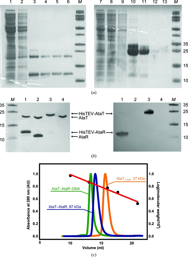
The ataRT operon from enteropathogenic Escherichia coli encodes a toxin-antitoxin (TA) module with a recently discovered novel toxin activity. This new type II TA module targets translation initiation for cell-growth arrest. Virtually nothing is known regarding the molecular mechanisms of neutralization, toxin catalytic action or translation autoregulation. Here, the production, biochemical analysis and crystallization of the intrinsically disordered antitoxin AtaR, the toxin AtaT, the AtaR-AtaT complex and the complex of AtaR-AtaT with a double-stranded DNA fragment of the operator region of the promoter are reported. Because they contain large regions that are intrinsically disordered, TA antitoxins are notoriously difficult to crystallize. AtaR forms a homodimer in solution and crystallizes in space group P6122, with unit-cell parameters a = b = 56.3, c = 160.8 Å. The crystals are likely to contain an AtaR monomer in the asymmetric unit and diffracted to 3.8 Å resolution. The Y144F catalytic mutant of AtaT (AtaTY144F) bound to the cofactor acetyl coenzyme A (AcCoA) and the C-terminal neutralization domain of AtaR (AtaR44-86) were also crystallized. The crystals of the AtaTY144F-AcCoA complex diffracted to 2.5 Å resolution and the crystals of AtaR44-86 diffracted to 2.2 Å resolution. Analysis of these structures should reveal the full scope of the neutralization of the toxin AtaT by AtaR. The crystals belonged to space groups P6522 and P3121, with unit-cell parameters a = b = 58.1, c = 216.7 Å and a = b = 87.6, c = 125.5 Å, respectively. The AtaR-AtaT-DNA complex contains a 22 bp DNA duplex that was optimized to obtain high-resolution data based on the sequence of two inverted repeats detected in the operator region. It crystallizes in space group C2221, with unit-cell parameters a = 75.6, b = 87.9, c = 190.5 Å. These crystals diffracted to 3.5 Å resolution.
Keywords: AtaR; AtaT; Escherichia coli; acetyltransferase; protein–DNA complexes; toxin–antitoxin.
Figures


Similar articles
-
Mechanism of regulation and neutralization of the AtaR-AtaT toxin-antitoxin system.Nat Chem Biol. 2019 Mar;15(3):285-294. doi: 10.1038/s41589-018-0216-z. Epub 2019 Feb 4. Nat Chem Biol. 2019. PMID: 30718814
-
Crystal Structure of the Enterohemorrhagic Escherichia coli AtaT-AtaR Toxin-Antitoxin Complex.Structure. 2019 Mar 5;27(3):476-484.e3. doi: 10.1016/j.str.2018.11.005. Epub 2019 Jan 3. Structure. 2019. PMID: 30612860
-
Crystallization and preliminary X-ray analysis of two variants of the Escherichia coli O157 ParE2-PaaA2 toxin-antitoxin complex.Acta Crystallogr F Struct Biol Commun. 2014 Sep;70(Pt 9):1284-91. doi: 10.1107/S2053230X1401749X. Epub 2014 Aug 27. Acta Crystallogr F Struct Biol Commun. 2014. PMID: 25195911 Free PMC article.
-
Novel toxins from type II toxin-antitoxin systems with acetyltransferase activity.Plasmid. 2017 Sep;93:30-35. doi: 10.1016/j.plasmid.2017.08.005. Epub 2017 Sep 20. Plasmid. 2017. PMID: 28941941 Review.
-
Keeping the Wolves at Bay: Antitoxins of Prokaryotic Type II Toxin-Antitoxin Systems.Front Mol Biosci. 2016 Mar 22;3:9. doi: 10.3389/fmolb.2016.00009. eCollection 2016. Front Mol Biosci. 2016. PMID: 27047942 Free PMC article. Review.
Cited by
-
Small-Molecule Acetylation by GCN5-Related N-Acetyltransferases in Bacteria.Microbiol Mol Biol Rev. 2020 Apr 15;84(2):e00090-19. doi: 10.1128/MMBR.00090-19. Print 2020 May 20. Microbiol Mol Biol Rev. 2020. PMID: 32295819 Free PMC article. Review.
-
Bacterial type II toxin-antitoxin systems acting through post-translational modifications.Comput Struct Biotechnol J. 2020 Dec 11;19:86-93. doi: 10.1016/j.csbj.2020.12.002. eCollection 2021. Comput Struct Biotechnol J. 2020. PMID: 33384857 Free PMC article. Review.
References
Publication types
MeSH terms
Substances
Grants and funding
- F.4505.16/Fonds De La Recherche Scientifique - FNRS/International
- U.N043.17F/Fonds De La Recherche Scientifique - FNRS/International
- CR-2017S-03/Fonds De La Recherche Scientifique - FNRS/International
- T.0147.15F PDR/Fonds De La Recherche Scientifique - FNRS/International
- J.0061.16F CDR/Fonds De La Recherche Scientifique - FNRS/International
LinkOut - more resources
Full Text Sources
Other Literature Sources
Research Materials
