. 2018 Jul;22(4):1168-1176.
doi: 10.1109/JBHI.2017.2762520.

Optimized Computer-Aided Segmentation and Three-Dimensional Reconstruction Using Intracoronary Optical Coherence Tomography

Optimized Computer-Aided Segmentation and Three-Dimensional Reconstruction Using Intracoronary Optical Coherence Tomography

Lambros Athanasiou et al. IEEE J Biomed Health Inform. 2018 Jul.

Abstract

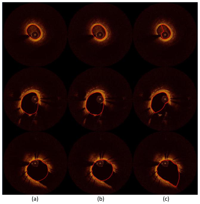
We present a novel and time-efficient method for intracoronary lumen detection, which produces three-dimensional (3-D) coronary arteries using optical coherence tomographic (OCT) images. OCT images are acquired for multiple patients and longitudinal cross-section (LOCS) images are reconstructed using different acquisition angles. The lumen contours for each LOCS image are extracted and translated to 2-D cross-sectional images. Using two angiographic projections, the centerline of the coronary vessel is reconstructed in 3-D, and the detected 2-D contours are transformed to 3-D and placed perpendicular to the centerline. To validate the proposed method, 613 manual annotations from medical experts were used as gold standard. The 2-D detected contours were compared with the annotated contours, and the 3-D reconstructed models produced using the detected contours were compared to the models produced by the annotated contours. Wall shear stress (WSS), as dominant hemodynamics factor, was calculated using computational fluid dynamics and 844 consecutive 2-mm segments of the 3-D models were extracted and compared with each other. High Pearson's correlation coefficients were obtained for the lumen area (r = 0.98) and local WSS (r = 0.97) measurements, while no significant bias with good limits of agreement was shown in the Bland-Altman analysis. The overlapping and nonoverlapping areas ratio between experts' annotations and presented method was 0.92 and 0.14, respectively. The proposed computer-aided lumen extraction and 3-D vessel reconstruction method is fast, accurate, and likely to assist in a number of research and clinical applications.


Figures

Fig. 1
Fig. 1
(a) The polar 2D data grayscale image (A-lines image), (b) its corresponding Cartesian 2D cross-sectional image, (c) the cross-section lines of the four different angles (0°, 45°, 90° and 135°) in the 2D data grayscale image and (d) its corresponding 2D cross-sectional image.
Fig. 2
Fig. 2
The four constructed LOCS images one for each angle: 0°, 45°, 90° and 135° (grayscale images) using all the 2D cross-sectional images (colored images-top).
Fig. 3
Fig. 3
The lumen detection in a LOCS image: (a) initial LOCS image, (b) filtered LOCS image, (c) segmented using K-means LOCS image showing the scan direction for the detection of non-zero pixels and (d) the connected nonzero pixels which represent the LOCS lumen borders.
Fig. 4
Fig. 4
(a) The four different LOCS images, their corresponding detected lumen borders (white) and the n (n ∈ [0 N]) row of the LOCS images corresponding to the n frame of the OCT pullback and to one of the four angles (0°-yellow row, 45°-green row, 90°-red row and 135°-white row). (b) The nth 2D OCT image having 8 points which correspond to nth the row of each LOCS image. (c) The cubic spline function applied to the 8 points representing the lumen border of the 2D OCT image.
Fig. 5
Fig. 5
(a) Correlation plot and (b) Bland-Altman plots for the lumen areas estimated by our method and experts annotations.
Fig. 6
Fig. 6
Volume comparison of the 3D arteries produced using expert annotations and algorithm’s lumen detection.
Fig. 7
Fig. 7
(a) Correlation plot and (b) Bland-Altman plots for the wall shear stress estimated by our method and experts.
Fig. 8
Fig. 8
Representative three-dimensional (3D) reconstructed models using the annotated (middle) and the proposed method detected lumen contours (right). The models are color-coded by wall shear stress (WSS) values (left).
Fig. 9
Fig. 9
Histogram with a Gaussian fit for the difference in lumen areas between the annotations - proposed method (green) and the annotations - literature [22] method (blue).
Fig. 10
Fig. 10
Ability of the proposed method on correcting existing methods drawbacks: three-dimensional (3D) reconstructed model using (a) the annotated, (b) the proposed method, and (c) the literature method [22] lumen contours highlighted with red color on the OCT images. The literature method [22] fails to detect the lumen contour in areas having artifacts (d): the presented OCT image (a, b, c: top and d) has residual blood inside the catheter tip and the lumen area.
Fig. 11
Fig. 11
Application examples of the proposed method and literature method showing the ability of our method in detecting more accurately the lumen border in segments having branches (middle and bottom image sets) and residual blood artifacts (top image set): (a) annotated images, (b) the result of the proposed method, and (c) the result of the literature’s method [22]. The lumen contours are highlighted with red.

References

    1. Stone GW, et al. A prospective natural-history study of coronary atherosclerosis. N Engl J Med. 2011;364(3):226–235. doi: 10.1056/NEJMoa1002358. - DOI - PubMed
    1. Athanasiou LS, Fotiadis DI, Michalis LK. Atherosclerotic Plaque Characterization Methods Based on Coronary Imaging. Elsevier Science; 2017.
    1. Regar E, van Leeuwen AMG, Serruys P. Optical Coherence Tomography in Cardiovascular Research. United Kingdom: Informa Healthcare; 2007.
    1. McCabe JM, Croce KJ. Optical coherence tomography. Circulation. 2012;126(17):2140–2143. - PubMed
    1. Bezerra HG, Costa MA, Guagliumi G, Rollins AM, Simon DI. Intracoronary Optical Coherence Tomography: A Comprehensive Review Clinical and Research Applications. Jacc-Cardiovascular Interv. 2009;2(11):1035–1046. - PMC - PubMed

Publication types

MeSH terms