Review
. 2018 Aug:200:43-51.
doi: 10.1016/j.imlet.2018.06.009. Epub 2018 Jun 30.

Taking the lead - how keratinocytes orchestrate skin T cell immunity

Affiliations
Review

Taking the lead - how keratinocytes orchestrate skin T cell immunity

M M Klicznik et al. Immunol Lett. 2018 Aug.

Abstract

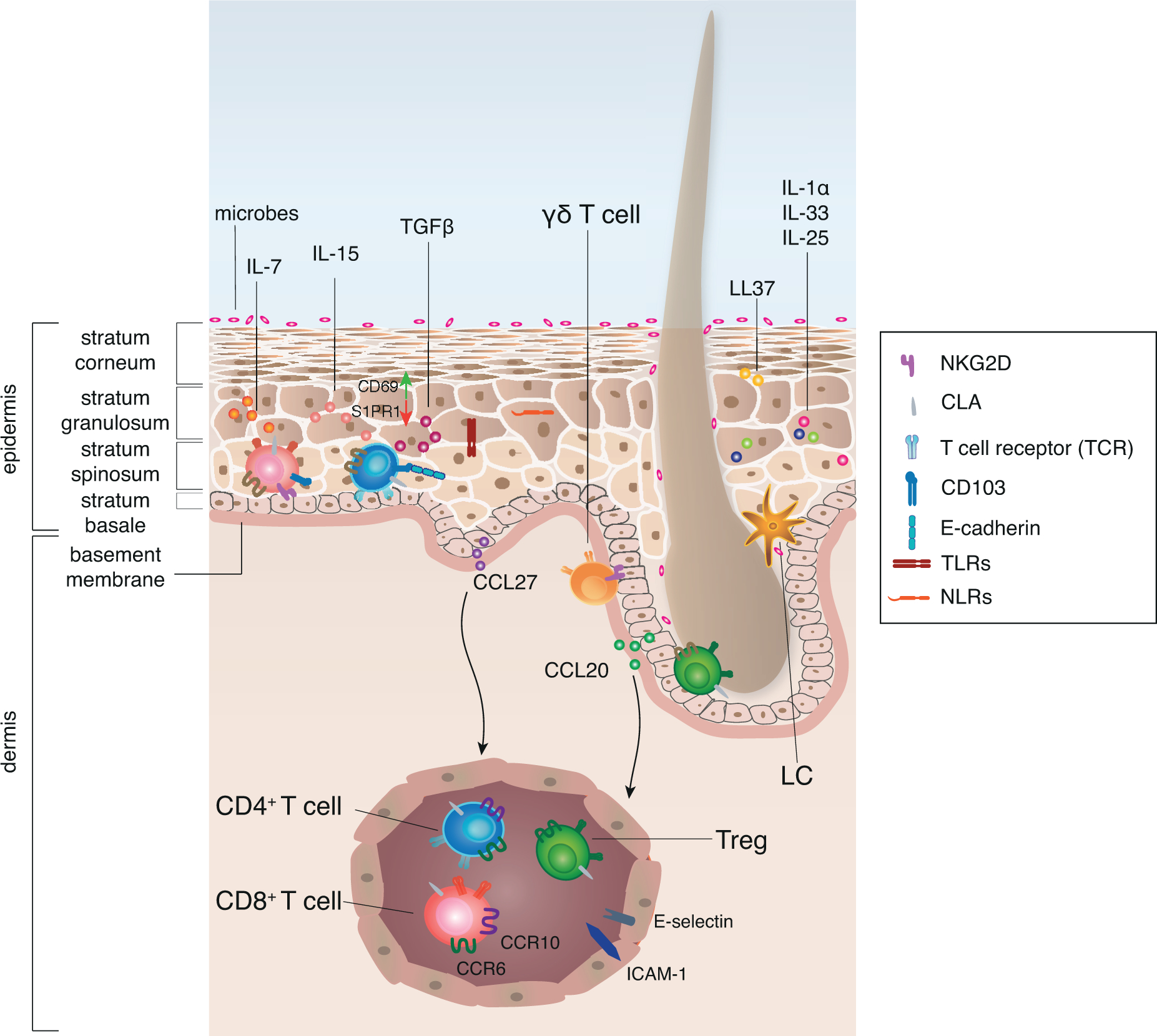
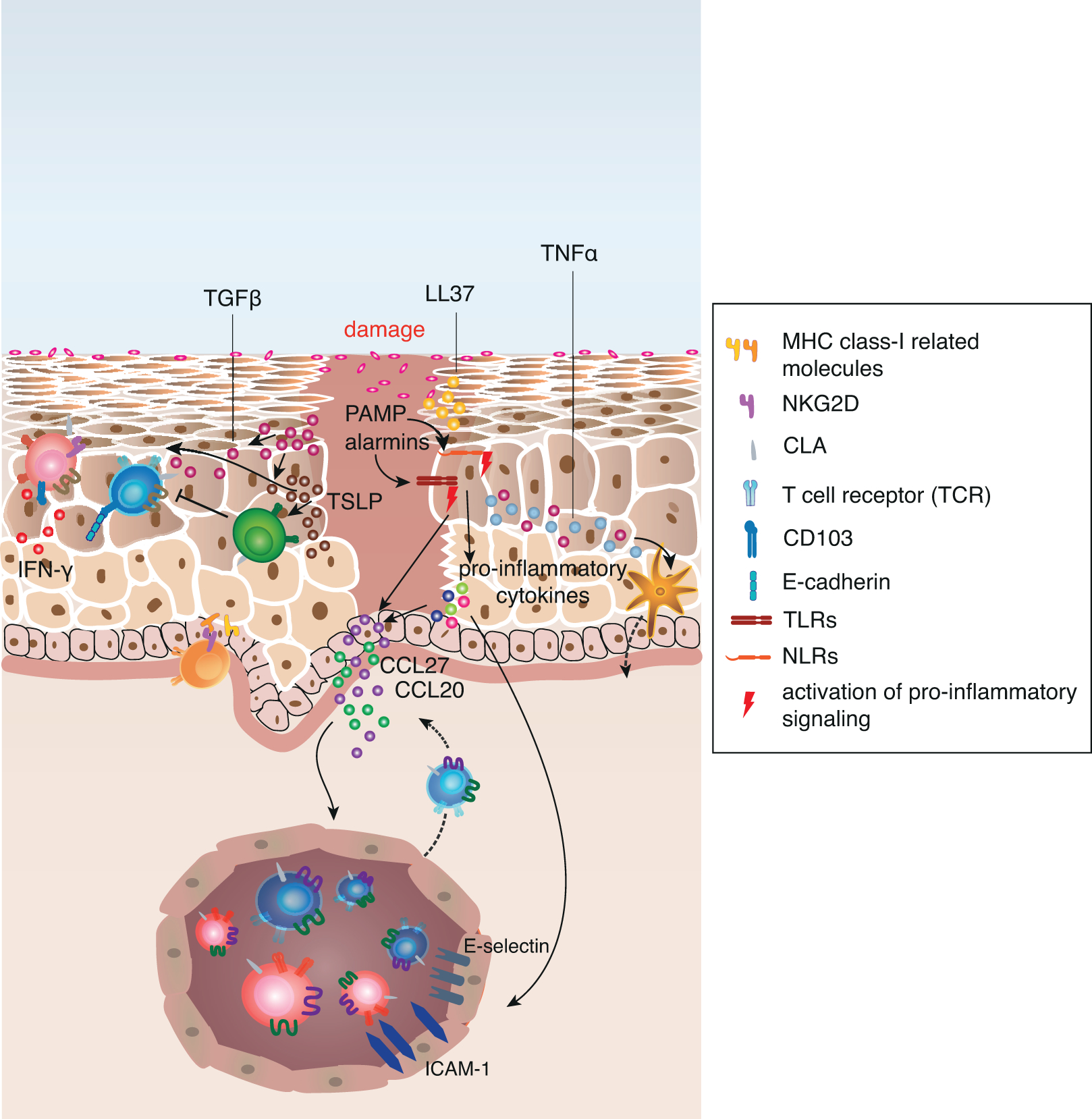
The skin comprises a complex coordinated system of epithelial tissue cells and immune cells that ensure adequate immune reactions against trauma, toxins and pathogens, while maintaining tissue homeostasis. Keratinocytes form the outermost barrier of the skin, and sense changes in barrier integrity, intrusion of microbial components and stress molecules. Thus, they act as sentinels that continuously communicate the status of the organ to the cutaneous immune system. Upon damage the keratinocytes initiate a pro-inflammatory signaling cascade that leads to the activation of resident immune cells. Simultaneously, the tissue mediates and supports immune-suppressive functions to contain inflammation locally. After resolution of inflammation, the skin provides a niche for regulatory and effector memory T cells that can quickly respond to reoccurring antigens. In this review we discuss the central role of keratinocyte-derived signals in controlling cutaneous T cell immunity.


Conflict of interest statement

Declarations of interest: none

Figures

Figure 1:
Figure 1:. Keratinocytes are equipped to sense tissue status and maintain T cell niche.
The human epidermis is mainly comprised of keratinocytes and can be divided into different layers. Keratinocytes function as sentinels that are equipped with TLRs and NLRs to sense danger signals, such as microbial components, chemicals or stress signals released upon damage, so called PAMPs and alarmins. Alarmins are produced by keratinocytes themselves and show anti-microbial properties, such as secretion of LL37. Additionally, keratinocytes are equipped with pre-stored cytokines, such as IL-1α, IL-25 and IL-33 that are released upon cell damage. The immune function of the epidermis is supported by specialized populations of antigen-presenting cells, CD1a+ Langerhans cells (LC), as well as innate lymphoid cells, and adaptive tissue-resident T cells. CCL20 produced by keratinocytes of the HF co-localizes LC and Treg and thus HF may serve as a niche for the induction of peripheral tolerance. Similarly, γδ T cells are often associated with adnexal structures. The human skin provides a variety of survival and maintenance signals such as IL-7, IL-15 and TGF-β to support the maintenance of tissue-resident T cells that ensure quick response to infection. Keratinocyte-derived CCL27 and CCL20 allow for the recruitment of T cells from the blood that express CCR10 and CCR6 respectively to non-inflamed tissue. T cells that sense the chemokine gradient can extravasate through post-capillary venules mediated by E-selectin - CLA interaction and ICAM-1. Upon tissue entry T cells change their surface receptor expression, probably induced by tissue-derived signals. In order to maintain antigen-experienced T cells within the epidermis, downregulation of S1PR1 and upregulation of CD69 are supported by IL-15 and TGF-β. In addition, TGF-β induces upregulation of CD103 on T cells that allows them to interact with E-cadherin on keratinocytes, which supports the retention of resident memory T cells.
Figure 2:
Figure 2:. Keratinocytes orchestrate damage response and its regulation in human skin.
Upon tissue damage pre-stored cytokines are released from damaged cells and initiate immediate effector functions in adjacent cells. Sensing of microbial components and stress molecules (PAMPs and alarmins) by TLRs and NLRs result in the activation of pro-inflammatory signalling pathways such as NF-κB and IRF transcription. This leads to the increased production of pro-inflammatory cytokines, such as IL-1α, IL-1β, IL-18, TNFα and type I interferons. Keratinocyte-derived TNFα activates Langerhans cells to migrate to skin-draining lymph nodes and activate naive or central memory T cells that upregulate CLA expression upon interaction with the LC. Additionally, the pro-inflammatory cytokines mediate the upregulation of chemokines and adhesion molecules on post-capillary venules as well as on adjacent keratinocytes, which promotes recruitment of circulating skin-tropic T cells from the blood. In addition to these mechanisms, keratinocytes upregulate MHC class I-related gene products on their surface that engage NKG2D on CD8+ T cells and γ5 T cells. Pro-inflammatory cytokines also allow keratinocytes to directly activate antigen-experienced memory CD4+ T cells. Activated T cells in the tissue secrete pro-inflammatory cytokines, (e.g. IFN-γ), that in turn activate chemokine secretion by keratinocytes and increased alarmin and anti-microbial peptide (e.g. LL37) production. TGF-β and TSLP secretion is increased upon tissue damage, and while both cytokines induce a variety of pro-inflammatory gene products and induce migration of LCs to skin-draining lymph nodes, they are essential to keep the inflammation local and support its resolution by directly signaling to Treg. TGF-β also promotes the local formation of resident memory T cells to provide quick response in case of a secondary antigen encounter.

References

    1. Coskun M, Intestinal epithelium in inflammatory bowel disease, Front. Med 1 (2014) 24 10.3389/fmed.2014.00024. - DOI - PMC - PubMed
    1. Gandhi VD, Vliagoftis H, Airway epithelium interactions with aeroallergens: role of secreted cytokines and chemokines in innate immunity, Front. Immunol 6 (2015) 147 10.3389/fimmu.2015.00147. - DOI - PMC - PubMed
    1. Peterson LW, Artis D, Intestinal epithelial cells: regulators of barrier function and immune homeostasis, Nat. Rev. Immunol 14 (2014) 141–153. 10.1038/nri3608. - DOI - PubMed
    1. Kolarsick PAJ, Kolarsick MA, Goodwin C, Anatomy and Physiology of the Skin, J. Dermatol. Nurses Assoc 3 (2011) 203 10.1097/JDN.0b013e3182274a98. - DOI
    1. Baroni A, Buommino E, De Gregorio V, Ruocco E, Ruocco V, Wolf R, Structure and function of the epidermis related to barrier properties, Clin. Dermatol 30 (2012) 257–262. 10.1016/j.clindermatol.2011.08.007. - DOI - PubMed

Publication types

LinkOut - more resources