

Effect of different delivery modes on the short-term strength of the pelvic floor muscle in Chinese primipara
- PMID: 29970030
- PMCID: PMC6029267
- DOI: 10.1186/s12884-018-1918-7
Effect of different delivery modes on the short-term strength of the pelvic floor muscle in Chinese primipara
Abstract
Background: To investigate the effect of different delivery modes and related obstetric factors on the short-term strength of the pelvic floor muscle after delivery in Chinese primipara.
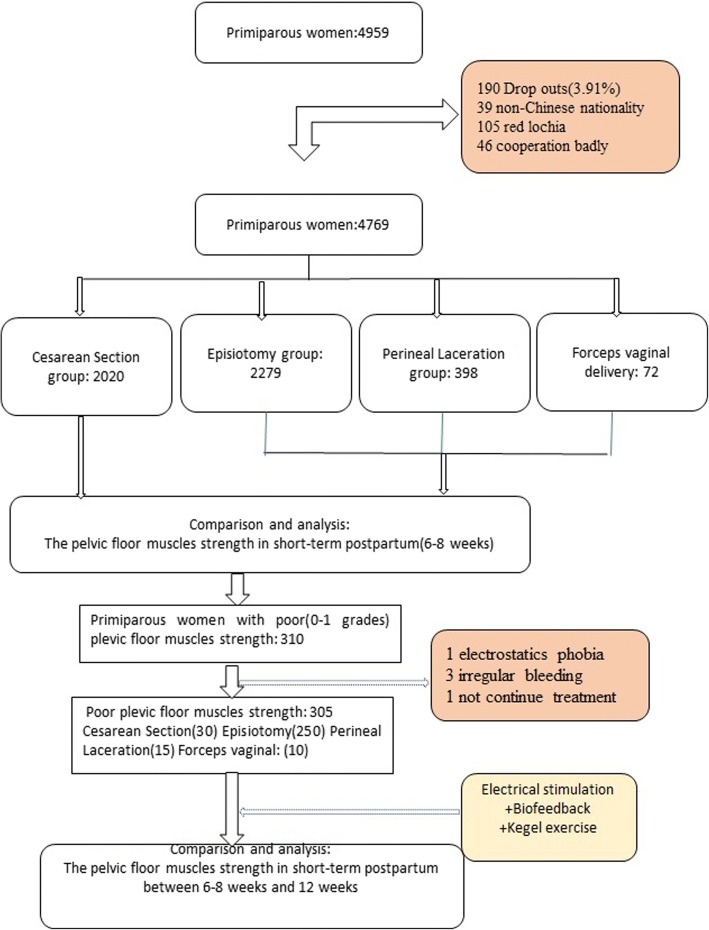
Methods: A total of 4769 healthy Chinese primiparas at postpartum 6-8 weeks were interviewed. According to the difference of delivery mode, the selected primiparas were divided into 2 groups, including cesarean delivery group containing 2020 and vaginal delivery group containing 2749. All the vaginal deliveries were further divided into 3 groups, including episiotomy group containing 2279, perineal laceration group containing 398, and forceps assisted group containing72. The scales of their pelvic floor muscle (PFM) strengths were examined by specially trained personnel using digital palpation (Modified Oxford scale:0-5 grade). According to participants' willingness, if the PFM strength was weak (0 or 1 grade), at-home PFM training would be recommended and an electrical stimulation combined with biofeedback therapy would be conducted for them in hospital. Twelve weeks after delivery, the PFM strength would be measured again. For statistical analysis, t-test, one-way variance analysis, Chi-square analysis, Kruskal-Wallis test H, Mann-Whitney U test and Wilcoxon test were carried out.
Results: The PFM strength in cesarean delivery group was higher than in vaginal delivery group (p < 0.05). Among 3 vaginal delivery groups, the PFM strength in perineal laceration group was the highest (p < 0.05); however, there was no difference in PFM strength between episiotomy group and forceps assisted group (p>0.05). After accepting PFM training at home and therapy in hospital, 305 women showed increased PFM strength (p < 0.05).
Conclusions: Vaginal delivery is an independent risk factor causing the damage of PFM, and episiotomy may cause injury of PFM. Through PFM training at home and therapy in hospital, those damage will resume as soon as possible in the short-time period after delivery.
Keywords: Biofeedback; Cesarean delivery; Eletrical stimulation; Episiotomy; Forceps; Pelvic floor muscle strength; Perineal laceration.
Conflict of interest statement
Ethics approval and consent to participate
The study protocol was approved by the Ethics Committee of Maternal and Child Health Hospital of Hubei Province(201301) and all included women signed written informed consent.
Consent for publication
Not applicable.
Competing interests
The authors declare that they have no competing interests.
Publisher’s Note
Springer Nature remains neutral with regard to jurisdictional claims in published maps and institutional affiliations.
Figures
Similar articles
-
The effect of water immersion delivery on the strength of pelvic floor muscle and pelvic floor disorders during postpartum period: An experimental study.Medicine (Baltimore). 2017 Oct;96(41):e8124. doi: 10.1097/MD.0000000000008124. Medicine (Baltimore). 2017. PMID: 29019880 Free PMC article.
-
Are there differences in short-term pelvic floor muscle function after cesarean section or vaginal delivery in primiparous women? A systematic review with meta-analysis.Int Urogynecol J. 2020 Aug;31(8):1497-1506. doi: 10.1007/s00192-020-04231-6. Epub 2020 Feb 15. Int Urogynecol J. 2020. PMID: 32062680
-
A Mobile Application Penyikang Applied in Postpartum Pelvic Floor Dysfunction: A Cross-Sectional Study to Analyze the Factors Influencing Postpartum Pelvic Floor Muscle Strength and Women's Participation in Treatment.Biomed Res Int. 2020 Jul 28;2020:4218371. doi: 10.1155/2020/4218371. eCollection 2020. Biomed Res Int. 2020. PMID: 32775419 Free PMC article.
-
Clinical and ultrasonographic evaluation of the pelvic floor in primiparous women: a cross-sectional study.Int Urogynecol J. 2018 Oct;29(10):1543-1549. doi: 10.1007/s00192-018-3581-y. Epub 2018 Mar 6. Int Urogynecol J. 2018. PMID: 29508047
-
Pelvic floor dysfunction, and effects of pregnancy and mode of delivery on pelvic floor.Taiwan J Obstet Gynecol. 2014 Dec;53(4):452-8. doi: 10.1016/j.tjog.2014.08.001. Taiwan J Obstet Gynecol. 2014. PMID: 25510682 Review.
Cited by
-
Effects of Different Delivery Modes on Pelvic Floor Function in Parturients 6-8 Weeks after Delivery Using Transperineal Four-Dimensional Ultrasound.Dis Markers. 2022 May 18;2022:2334335. doi: 10.1155/2022/2334335. eCollection 2022. Dis Markers. 2022. PMID: 35634438 Free PMC article.
-
Association of the second birth mode of delivery and interval with maternal pelvic floor changes: a prospective cohort study.BMC Pregnancy Childbirth. 2024 Mar 7;24(1):178. doi: 10.1186/s12884-024-06366-6. BMC Pregnancy Childbirth. 2024. PMID: 38454330 Free PMC article.
-
Polycystic Ovary Syndrome and Pelvic Floor Dysfunction: A Narrative Review.Res Rep Urol. 2020 May 7;12:179-185. doi: 10.2147/RRU.S249611. eCollection 2020. Res Rep Urol. 2020. PMID: 32440514 Free PMC article. Review.
-
Vaginal palpation versus transabdominal ultrasound in the comprehension of pelvic floor muscle contraction after vaginal delivery: a randomised controlled trial.BMC Womens Health. 2021 Feb 6;21(1):53. doi: 10.1186/s12905-021-01203-w. BMC Womens Health. 2021. PMID: 33549078 Free PMC article. Clinical Trial.
-
The effect of a pelvic compression belt on postural stability in postpartum women.Sports Eng. 2025;28(2):34. doi: 10.1007/s12283-025-00516-5. Epub 2025 Jul 28. Sports Eng. 2025. PMID: 40772253 Free PMC article.
References
MeSH terms
Grants and funding
LinkOut - more resources
Full Text Sources
Other Literature Sources
Medical
