

IgG4-related ophthalmic disease involving extraocular muscles: case series
- PMID: 29970061
- PMCID: PMC6029167
- DOI: 10.1186/s12886-018-0819-x
IgG4-related ophthalmic disease involving extraocular muscles: case series
Abstract
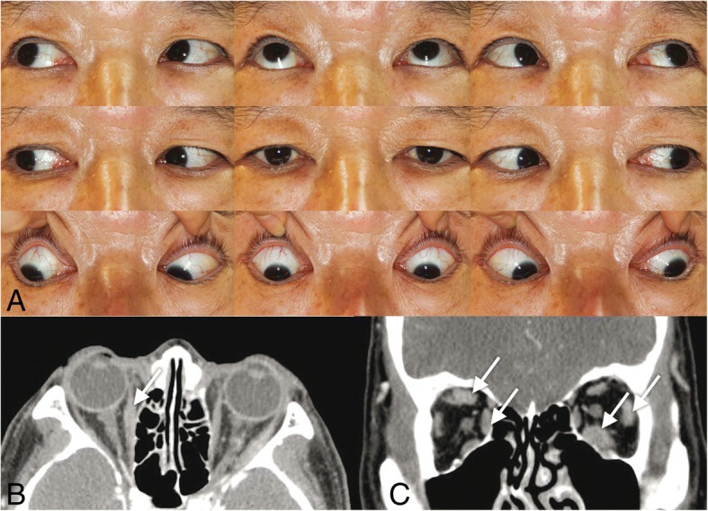
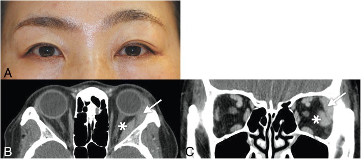
Background: To elucidate the clinical features of strabismus associated with IgG4-related ophthalmic disease (IgG4-ROD). All of the four patients with IgG4-ROD showed marked enlargement of the extraocular muscles, however, two patients showed orthotropia with full ductions and versions. One patient showed a small angle of exotropia and hypertropia of less than 5 prism diopters. One remaining patient showed orthotropia, full ductions and versions despite marked enlargement of the extraocular muscles, then developed hypertropia up to 35 prism diopters with activation of inflammation, which promptly improved after treatment with oral steroids.
Conclusions: IgG4-ROD usually shows normal ocular motility despite extraocular muscle enlargement, which is the key distinguishing feature from other orbital inflammatory diseases. Active flare-up with increased serum IgG4 levels may produce a large angle of eye deviation, but mostly respond well to steroid treatment.
Keywords: Case report; Extraocular muscle; IgG4-related ophthalmic disease; Imaging findings; Strabismus.
Conflict of interest statement
Ethics approval and consent to participate
This study complied with the tenets of the Declaration of Helsinki. This study received ethical approval from the Institutional Review Board of the Seoul National University Bundang Hospital.
Consent for publication
Written informed consent was obtained from the patients for publication and any accompanying images. A copy of the written consent is available for review by the Editor of this journal.
Competing interests
The authors declare that they have no competing interests.
Publisher’s Note
Springer Nature remains neutral with regard to jurisdictional claims in published maps and institutional affiliations.
Figures


Similar articles
-
Extraocular muscle surgery for extorsion after macular translocation surgery new surgical technique and clinical management.Ophthalmology. 2006 Jan;113(1):63-9. doi: 10.1016/j.ophtha.2005.09.022. Epub 2005 Nov 10. Ophthalmology. 2006. PMID: 16290047
-
Location and frequency of lesions in patients with IgG4-related ophthalmic diseases.Graefes Arch Clin Exp Ophthalmol. 2014 Mar;252(3):531-8. doi: 10.1007/s00417-013-2548-4. Epub 2014 Jan 3. Graefes Arch Clin Exp Ophthalmol. 2014. PMID: 24384801
-
Systemic Amyloidosis and Extraocular Muscle Deposition.J Neuroophthalmol. 2016 Jun;36(2):167-73. doi: 10.1097/WNO.0000000000000358. J Neuroophthalmol. 2016. PMID: 26967574
-
IgG4-Related Ophthalmic Disease. Part II: Clinical Aspects.Ophthalmic Plast Reconstr Surg. 2015 May-Jun;31(3):167-78. doi: 10.1097/IOP.0000000000000364. Ophthalmic Plast Reconstr Surg. 2015. PMID: 25564258 Review.
-
IgG4-related ophthalmic disease. Part I: background and pathology.Ophthalmic Plast Reconstr Surg. 2015 Mar-Apr;31(2):83-8. doi: 10.1097/IOP.0000000000000363. Ophthalmic Plast Reconstr Surg. 2015. PMID: 25564257 Review.
Cited by
-
Autoimmune disease of head and neck, imaging, and clinical review.Neuroradiol J. 2022 Oct;35(5):545-562. doi: 10.1177/19714009221100983. Epub 2022 May 22. Neuroradiol J. 2022. PMID: 35603923 Free PMC article. Review.
-
Normative measurements of the superior oblique and inferior oblique muscles by magnetic resonance imaging.Surg Radiol Anat. 2022 Apr;44(4):521-525. doi: 10.1007/s00276-022-02915-w. Epub 2022 Mar 8. Surg Radiol Anat. 2022. PMID: 35258651 Free PMC article.
-
Analysis of extraocular muscle volumes in idiopathic hypertrophic pachymeningitis patients.PLoS One. 2025 Apr 29;20(4):e0309638. doi: 10.1371/journal.pone.0309638. eCollection 2025. PLoS One. 2025. PMID: 40299826 Free PMC article.
-
Effects of Fibulin-5 Gene Silencing on Proliferation and Apoptosis of IgG4-ROD Lacrimal Gland Fibroblasts.Stem Cells Int. 2023 Feb 10;2023:2742839. doi: 10.1155/2023/2742839. eCollection 2023. Stem Cells Int. 2023. Retraction in: Stem Cells Int. 2023 Dec 20;2023:9856916. doi: 10.1155/2023/9856916. PMID: 36818161 Free PMC article. Retracted.
References
Publication types
MeSH terms
Substances
Grants and funding
LinkOut - more resources
Full Text Sources
Other Literature Sources
