. 2018 Jul 3;16(1):38.
doi: 10.1186/s12964-018-0248-8.

Peptides derived from the integrin β cytoplasmic tails inhibit angiogenesis

Affiliations

Peptides derived from the integrin β cytoplasmic tails inhibit angiogenesis

Zhongyuan Cao et al. Cell Commun Signal. .

Abstract

Background: Integrins are essential regulators of angiogenesis. However, the antiangiogenic potential of peptides derived from the integrin cytoplasmic tails (CT) remains mostly undetermined.

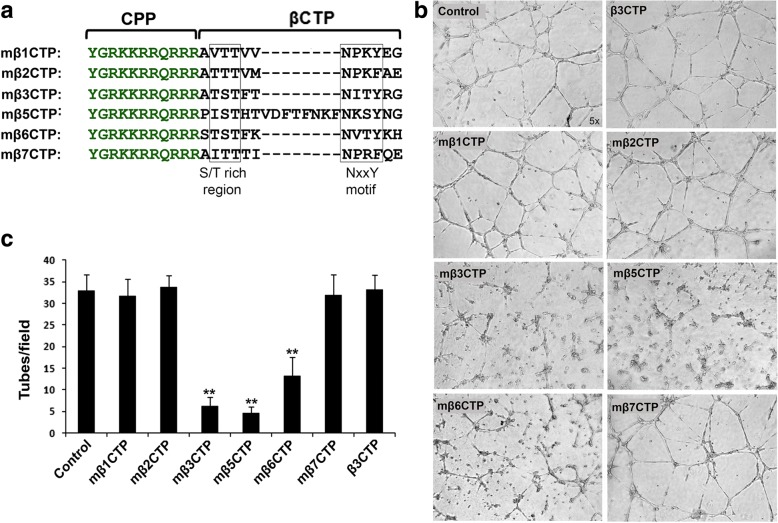
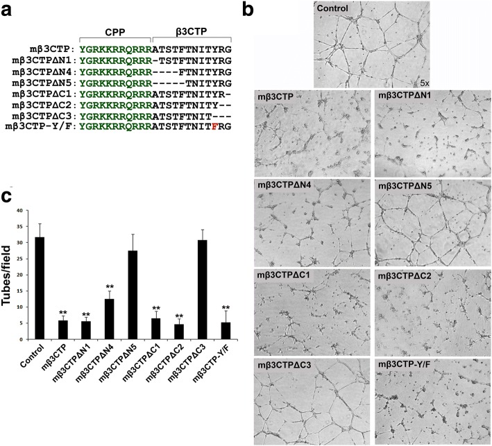
Methods: Here we designed a panel of membrane-penetrating peptides (termed as mβCTPs), each comprising a C-terminal NxxY motif from one of the conserved integrin β CTs, and evaluated their antiangiogenic ability using both in vitro and in vivo approaches.

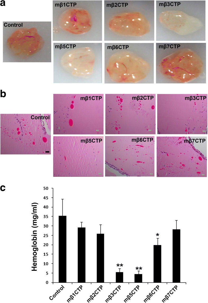
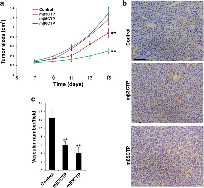
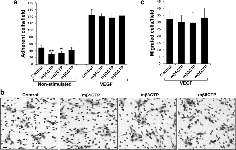
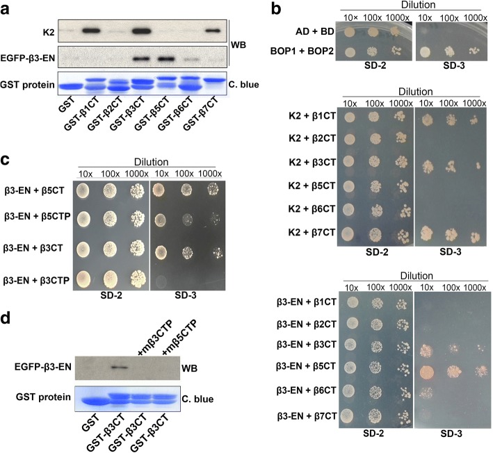
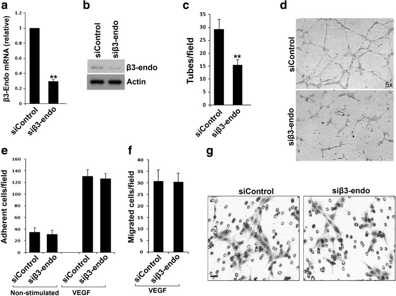
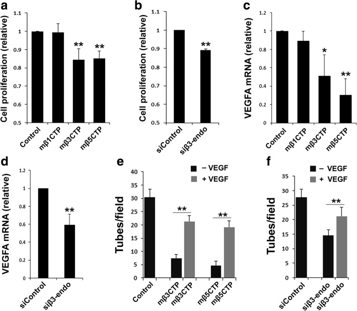
Results: We found that mβ3CTP, mβ5CTP and mβ6CTP, derived respectively from the integrin β3, β5 and β6 CTs, but not others, exhibit antiangiogenic ability. Interestingly, we observed that the integrin β3, β5 and β6 CTs but not others are able to interact with β3-endonexin. In addition, the antiangiogenic core in mβ3CTP is identical to a previously identified β3-endonexin binding region in the integrin β3 CT, indicating that the antiangiogenic mβCTPs may function via their binding to β3-endonexin. Consistently, knockdown of endogenous β3-endonexin in HUVECs significantly suppresses tube formation, suggesting that β3-endonexin is proangiogenic. However, neither treatment with the antiangiogenic mβCTPs nor knockdown of endogenous β3-endonexin affects integrin-mediated HUVEC adhesion and migration, indicating that their antiangiogenic effect may not rely on directly regulating integrin activity. Importantly, both treatment with the antiangiogenic mβCTPs and knockdown of endogenous β3-endonexin in HUVECs inhibit VEGF expression and cell proliferation, thereby providing mechanistic explanations for the functional consequences.

Conclusion: Our results suggest that the antiangiogenic mβCTPs can interact with β3-endonexin in vascular endothelial cells and suppress its function in regulating VEGF expression and cell proliferation, thus disclosing a unique pathway that may be useful for developing novel antiangiogenic strategies.

Keywords: Angiogenesis; Cytoplasmic tails; Endothelial cells; Integrins; β3-endonexin.


Conflict of interest statement

Ethics approval and consent to participate

All animal handling and experiments were approved by the IACUC of Shanghai University School of Life Sciences.

Consent for publication

Not applicable.

Competing interests

The authors declare that they have no competing interests.

Publisher’s Note

Springer Nature remains neutral with regard to jurisdictional claims in published maps and institutional affiliations.

Figures

Fig. 1
Fig. 1
The mβCTPs selectively inhibit HUVEC tube formation. a Amino-acid sequences of mβCTPs. In each of mβCTPs, a cell-penetrating peptide (CPP) was fused with a short amino-acid sequence from one of the C-termini of integrin β CTs (βCTP), comprising an NxxY/F motif and a proximal S/T cluster. b HUVECs were seeded on polymerized Matrigel with or without the indicated mβCTPs (20 μM at a final concentration) for 8 h. The formed capillary-like tube structures were recorded under an inverted microscope (5× objective). c The capillary-like polygon tubes were quantified. Results were expressed as means ±SD of three or more experiments, and statistical significance was analyzed using Student’s t test (**, p < 0.01)
Fig. 2
Fig. 2
The mβCTPs selectively suppress blood vessel formation in implanted Matrigel plugs. a Aliquots of Matrigel liquid supplemented with the indicated mβCTPs (50 μM at a final concentration) were subcutaneously injected into BALB/c nude mice (n = 5) to form solid Matrigel plugs. Matrigel that did not contain peptides was used as a control. After 7 days, Matrigel plugs were carefully isolated and photographed. b Matrigel plugs were fixed and paraffin-embedded for histological analysis. Representative H&E staining images of the Matrigel plug sections were shown. Scale bar, 200 μm. c The amount of hemoglobin in Matrigel plugs was quantified by Drakin’s reagent. Results were expressed as means ± SD of five samples; statistical significance was analyzed using Student’s t test (*, p < 0.05; **, p < 0.01)
Fig. 3
Fig. 3
The antiangiogenic mβCTPs show antitumor activity. a 1.2 × 106 of RM1 cancer cells were subcutaneously injected into BALB/c nude mice (n = 6). Starting on day 5, the formed tumor areas were subjected to treatment by local injection of 100 μl of the indicated mβCTP solution (50 μM at a final concentration) every other day. PBS alone was used as a control. Length (l) and width (w) of the formed tumors were measured, and tumor volumes (v) were calculated by using the following formula: v = 0.52(l × w2). b, c The tumor tissues were harvested on day 15 and subjected to IHC staining for CD31, a marker of vascular endothelial cells. Blood vessels in solid tumor tissues were quantified. Scale bar, 50 μm. The results were expressed as means ±SD; statistical significance was analyzed using Student’s t test (**, p < 0.01)
Fig. 4
Fig. 4
The antiangiogenic mβCTPs fail to affect VEGF-induced HUVEC adhesion and migration. a HUVECs were treated with the indicated mβCTPs (20 μM) and allowed to adhere to immobilized fibrinogen for 30 min in the absence or presence of 20 ng/ml of VEGF. The adherent cells were fixed, stained and counted as described in methods. b, c HUVECs were treated with the indicated mβCTPs (20 μM) and allowed to migrate to Transwell membrane coated with fibrinogen for 8 h in the presence of VEGF (20 ng/ml). The migrated cells were fixed, stained, photographed and counted. Scale bar, 20 μm. Results were expressed as means ± SD of five samples; statistical significance was analyzed using Student’s t test (*, p < 0.05; **, p < 0.01)
Fig. 5
Fig. 5
Identify the antiangiogenic core region in mβ3CTP. a The amino-acid sequences of different mβ3CTP variants with sequential residue deletions from either side of β3CTP were shown. b, c HUVECs were seeded on polymerized Matrigel in 24-well plates and treated with the indicated mβ3CTP variants (20 μM at a final concentration) or PBS alone as a control. The formed capillary-like structures were counted under an inverted microscope (5× objective). Results were expressed as means ±SD from three experiments; statistical significance was analyzed using Student’s t test (**, p < 0.01)
Fig. 6
Fig. 6
The antiangiogenic mβCTPs specifically interact with β3-endonexin. a Lysate aliquots of HUVECs exogenously expressing EGFP-β3-endonexin (EGFP-β3-EN) were incubated with purified GST or GST-fused β CT proteins coupled on Glutathione Sepharose beads. After incubation, the beads were washed and the precipitated proteins were separated by SDS-PAGE. The loaded GST proteins on the beads were assessed by Coomassie blue (C. blue) staining. The co-precipitated EGFP-β3-endonexin and endogenous kindlin-2 (K2) were detected by Western blotting. b Interaction of the β CTs with β3-EN or K2 was evaluated using the yeast 2-hybrid system by a serial dilution method on selection media. Two known interacting molecules (Bop1 and Bop2) were employed here as a positive control, and empty vectors were transformed to serve as a negative control. The growth of yeast cells on SD-2 media (−Leu/−Trp) indicates a successful transformation; the growth on SD-3 selection media (−Leu/−Trp/-His) indicates a positive protein-protein interaction. c The interaction of β3-EN with β3CTP and β5CTP was evaluated using the yeast 2-hybrid system. d Interaction between EGFP-β3-endonexin and GST-β3 CT was evaluated by a pull-down assay in the presence of mβ3CTP or mβ5CTP (20 μM at a final concentration) as described in a
Fig. 7
Fig. 7
β3-Endonexin is able to promote HUVEC tube formation but not required for VEGF-induced HUVEC adhesion and migration. a, b HUVECs were transfected with an siRNA duplex specifically targeting β3-endonexin (siβ3-endo). A non-targeting siRNA duplex (siControl) was used as a control. Cells were harvested 48 h after transfection, and expression of β3-endonexin in HUVECs was quantified by qRT-PCR for mRNA (a) and western blotting for protein (b). c, d After transfected with siRNA for 48 h, HUVECs were harvested and used for the tube formation assays. The formed capillary-like structures were counted and the disconnected structures were indicated (arrow heads). e-g HUVECs transfected with either siControl or siβ3-endo were used in adhesion (e) and migration (f, g) assays in the absence and presence of VEGF. The adhered or migrated cells were quantified as described in methods. Results were expressed as means ±SD from three experiments; statistical significance was analyzed using Student’s t test (**, p < 0.01)
Fig. 8
Fig. 8
Treatment with the antiangiogenic mβCTPs and knockdown of endogenous β3-endonexin in HUVECs both suppress cell proliferation and VEGFA expression. HUVECs were treated with the indicated mβCTPs (20 μM at a final concentration) or transfected with siRNA (siControl or siβ3-endo). a, b HUVEC proliferation was evaluated using CCK-8 cell proliferation assay. c, d The expression levels of VEGFA mRNA in HUVECs were quantified by qRT-PCR. e, f HUVECs were treated with either siRNAs or mβCTPs as indicated and seeded on polymerized Matrigel in 24-well plates in the absence or presence of VEGF (20 ng/ml). The formed capillary-like structures were counted under an inverted microscope (5× objective). Results were expressed as means ±SD of three experiments; statistical significance was analyzed using Student’s t test (*, p < 0.05; **, p < 0.01)

Similar articles

Cited by

References

    1. Hynes RO. Integrins: bidirectional, allosteric signaling machines. Cell. 2002;110:673–687. doi: 10.1016/S0092-8674(02)00971-6. - DOI - PubMed
    1. Ginsberg MH, Loftus JC, D'Souza S, Plow EF. Ligand binding to integrins: common and ligand specific recognition mechanisms. Cell Diff Dev. 1990;32:203–213. doi: 10.1016/0922-3371(90)90033-S. - DOI - PubMed
    1. Tucker GC. Integrins: molecular targets in cancer therapy. Curr Oncol Rep. 2006;8:96–103. doi: 10.1007/s11912-006-0043-3. - DOI - PubMed
    1. Avraamides CJ, Garmy-Susini B, Varner JA. Integrins in angiogenesis and lymphangiogenesis. Nat Rev Cancer. 2008;8:604–617. doi: 10.1038/nrc2353. - DOI - PMC - PubMed
    1. Atkinson SJ, Ellison TS, Steri V, Gould E, Robinson SD. Redefining the role(s) of endothelial alphavbeta3-integrin in angiogenesis. Biochem Soc Trans. 2014;42:1590–1595. doi: 10.1042/BST20140206. - DOI - PubMed

Publication types

MeSH terms

Substances

LinkOut - more resources