

Expression profiling of Trypanosoma congolense genes during development in the tsetse fly vector Glossina morsitans morsitans
- PMID: 29970164
- PMCID: PMC6029126
- DOI: 10.1186/s13071-018-2964-8
Expression profiling of Trypanosoma congolense genes during development in the tsetse fly vector Glossina morsitans morsitans
Abstract
Background: The tsetse transmitted parasitic flagellate Trypanosoma congolense causes animal African trypanosomosis (AAT) across sub-Saharan Africa. AAT negatively impacts agricultural, economic, nutritional and subsequently, health status of the affected populace. The molecular mechanisms that underlie T. congolense's developmental program within tsetse are largely unknown due to considerable challenges with obtaining sufficient parasite cells to perform molecular studies.
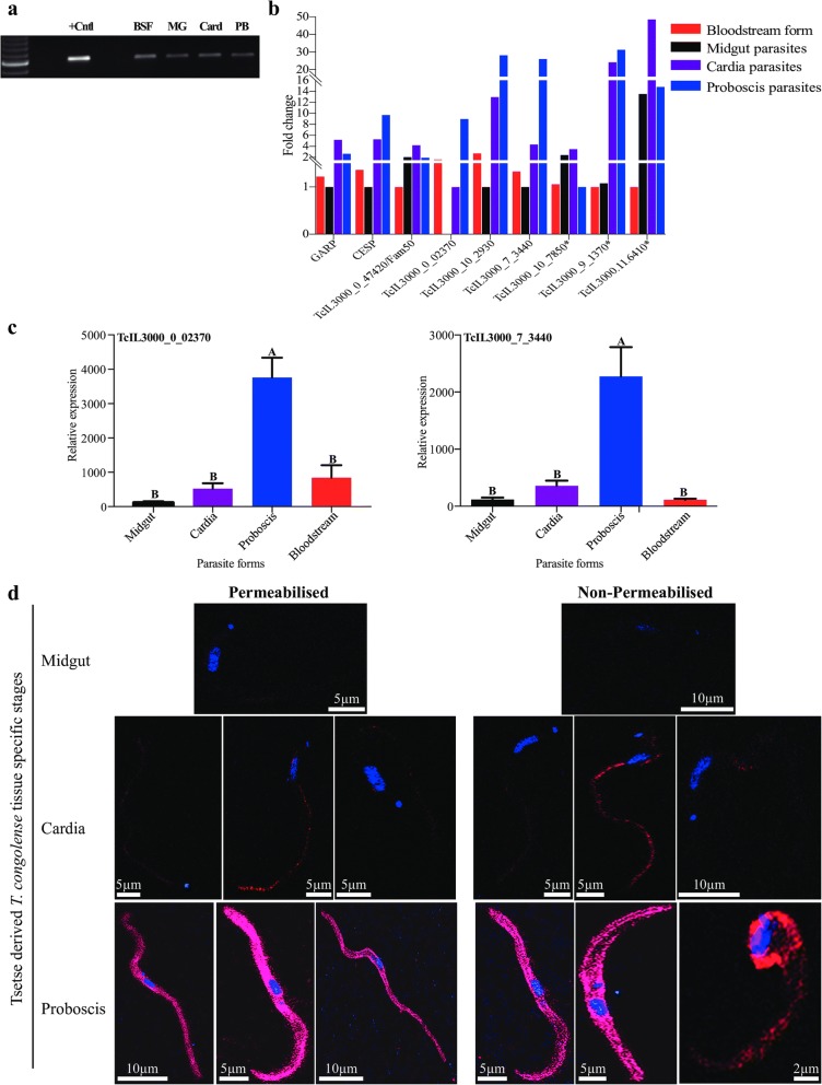
Methods: In this study, we used RNA-seq to profile T. congolense gene expression during development in two distinct tsetse tissues, the cardia and proboscis. Indirect immunofluorescent antibody test (IFA) and confocal laser scanning microscope was used to localize the expression of a putative protein encoded by the hypothetical protein (TcIL3000_0_02370).
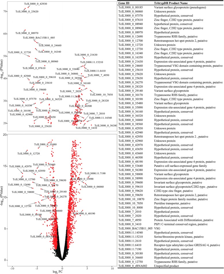
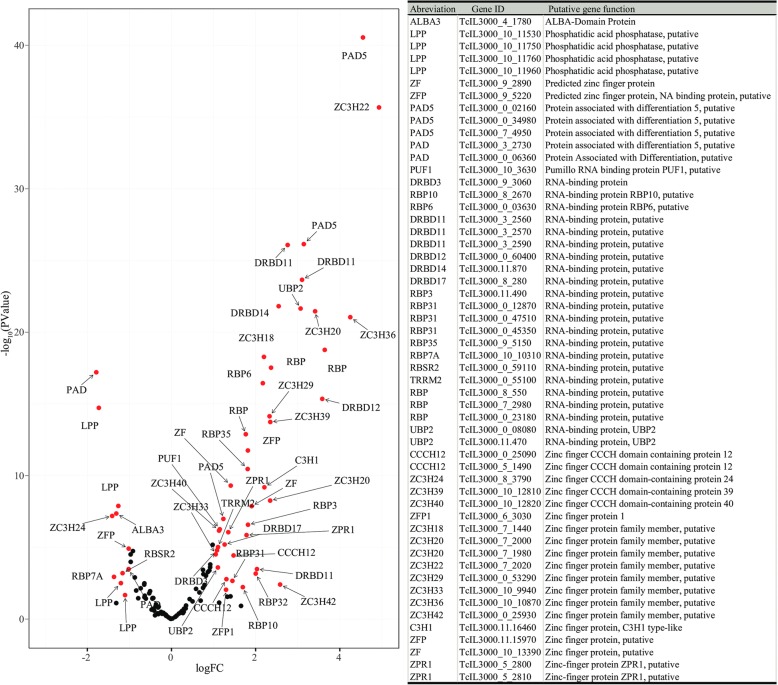
Results: Consistent with current knowledge, genes coding several variant surface glycoproteins (including metacyclic specific VSGs), and the surface coat protein, congolense epimastigote specific protein, were upregulated in parasites in the proboscis (PB-parasites). Additionally, our results indicate that parasites in tsetse's cardia (C-parasites) and PB employ oxidative phosphorylation and amino acid metabolism for energy. Several genes upregulated in C-parasites encoded receptor-type adenylate cyclases, surface carboxylate transporter family proteins (or PADs), transport proteins, RNA-binding proteins and procyclin isoforms. Gene ontology analysis of products of genes upregulated in C-parasites showed enrichment of terms broadly associated with nucleotides, microtubules, cell membrane and its components, cell signaling, quorum sensing and several transport activities, suggesting that the parasites colonizing the cardia may monitor their environment and regulate their density and movement in this tissue. Additionally, cell surface protein (CSP) encoding genes associated with the Fam50 'GARP', 'iii' and 'i' subfamilies were also significantly upregulated in C-parasites, suggesting that they are important for the long non-dividing trypomastigotes to colonize tsetse's cardia. The putative products of genes that were upregulated in PB-parasites were linked to nucleosomes, cytoplasm and membrane-bound organelles, which suggest that parasites in this niche undergo cell division in line with prior findings. Most of the CSPs upregulated in PB-parasites were hypothetical, thus requiring further functional characterization. Expression of one such hypothetical protein (TcIL3000_0_02370) was analyzed using immunofluorescence and confocal laser scanning microscopy, which together revealed preferential expression of this protein on the entire surface coat of T. congolense parasite stages that colonize G. m. morsitans' proboscis.
Conclusion: Collectively, our results provide insight into T. congolense gene expression profiles in distinct niches within the tsetse vector. Our results show that the hypothetical protein TcIL3000_0_02370, is expressed on the entire surface of the trypanosomes inhabiting tsetse's proboscis. We discuss our results in terms of their relevance to disease transmission processes.
Keywords: Gene expression analysis; Glossina morsitans morsitans; Trypanosoma congolense; Tsetse cardia; Tsetse proboscis and confocal microscopy.
Conflict of interest statement
Ethics approval
Animal experiments were carried out in strict accordance with recommendations in the Office of Laboratory Animal Welfare at the National Institutes of Health and Yale University Institutional Animal Care and Use Committee (ACUC). The experimental protocol was reviewed and approved by the Yale University Institutional Animal Care and Use Committee (Protocol number 2014-07266).
Consent for publication
Not applicable
Competing interests
The authors declared that they have no competing interests.
Publisher’s Note
Springer Nature remains neutral with regard to jurisdictional claims in published maps and institutional affiliations.
Figures


References
-
- Mattioli RC, Hendrickx G, Wint W, Jannin J, Slingenbergh JFU. Tsetse and trypanosomiasis intervention policies supporting sustainable animal-agricultural development. Food Agric Environ. 2004;2:310–412.
-
- Maudlin I, Holmes PH, Miles MA. The trypanosomiasis. Wallingford, UK: CAB International; 2004.
MeSH terms
Substances
Grants and funding
LinkOut - more resources
Full Text Sources
Other Literature Sources
Research Materials
Miscellaneous
