. 2018 Oct;17(10):1991-2004.
doi: 10.1074/mcp.RA118.000745. Epub 2018 Jul 3.

Proteomic Profiling of Microtubule Self-organization in M-phase

Affiliations

Proteomic Profiling of Microtubule Self-organization in M-phase

Miquel Rosas-Salvans et al. Mol Cell Proteomics. 2018 Oct.

Abstract

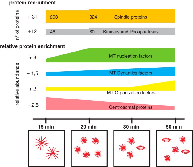
Microtubules (MTs) and associated proteins can self-organize into complex structures such as the bipolar spindle, a process in which RanGTP plays a major role. Addition of RanGTP to M-phase Xenopus egg extracts promotes the nucleation and self-organization of MTs into asters and bipolar-like structures in the absence of centrosomes or chromosomes. We show here that the complex proteome of these RanGTP-induced MT assemblies is similar to that of mitotic spindles. Using proteomic profiling we show that MT self-organization in the M-phase cytoplasm involves the non-linear and non-stoichiometric recruitment of proteins from specific functional groups. Our study provides for the first time a temporal understanding of the protein dynamics driving MT self-organization in M-phase.

Keywords: Cell biology; Cell division; Microtubule; Mitosis; Protein complex analysis; Protein-Protein Interactions; Proteomic profiling; RanGTP; Self-organization; Spindle.


Figures

Fig. 1.
Fig. 1.
RanGTP-induced MT self-organization in Xenopus egg extracts. A, Representative fluorescence images of RanGTP induced MT assemblies in egg extract upon incubation with RanGTP. Scale bar = 20 μm. B, Quantification of the different types of MT assemblies in 1 μl of egg extract at different times of incubation as indicated. Red bars, MT asters with long MTs; Orange bars, MT asters with short MTs; Blue bars, minispindles; Brown bars, aggregates of MT assemblies. Data obtained from four independent experiments. C, Colloidal blue stained SDS-PAGE of the MT pellets obtained at different time of incubation of egg extracts with RanGTP (as indicated). Molecular weight markers are indicated on the left in kDa. D, Schematic representation of the conversion and filtering process of the Xenopus proteins identified by mass spectrometry to obtain the final RanMT proteome of human proteins.
Fig. 2.
Fig. 2.
The RanMT proteome. A, Distribution of the GO terms of the RanMT proteome obtained with the online CleverGO tool (analysis conditions: Similarity strength threshold, 0.75; Minimal precision, 0.5; Minimal Lvl, 4; p value cutoff, 0.01). GO terms (represented as dots) more closely related are connected with a gray line. Four major clusters include more than 8 GO terms. The cluster that contains GO terms related to spindles is highlighted in red. B, Word cloud showing the words represented at the GO terms included in the cluster shown in red in (A). Word size is proportional to its frequency in the cluster GO terms. C, Venn-diagram showing the number of proteins identified in our study (RanMT blue) and the overlaps with the HeLa spindle proteomes described by Sauer et al. (8) (pink) and Rao et al. (10) (green) and the MeMT proteome described by Gache et al. (11) (gray). D, Bar graph showing the number of spindle proteins identified in our study (RanMT Sp. Prot., orange bar) and the overlaps with MiCroKiTS (dark gray bar), the HeLa spindle proteomes described by Sauer et al. (8) (magenta bar) and Rao et al. (10) (green bar) and the MeMT proteome described by Gache et al. (11) (brown bar). The number of spindle proteins that are unique to our study is shown on the right (Only RanMT, light gray bar).
Fig. 3.
Fig. 3.
Identification of novel SAFs. A, Immunofluorescence images from HeLa cells showing the localization of three proteins identified by mass spectrometry, CBX3, CK2, and DnaJB6 as indicated. Upper row, Immunofluorescence signal detected with antibodies against each of the proteins as indicated. Lower row, overlaid images (in green, signal for each protein as shown above, in red tubulin, and in blue DNA). Scale bar = 5 μm. B, Quantifications of MT regrowth assays in HeLa cells for Control and CBX3 or DnaJB6 silenced cells and for cells incubated with DMSO or with the CK2 inhibitor Quinalizarin (QZ) 30 μm. The graphs show the number of MT asters in mitotic cells fixed 3 min after nocodazole washout. n = 3, experiments performed in different days (total sample size, 413 control and 409 CBX3 silenced cells; 308 control DMSO and 323 QZ incubated cells; 220 control and 223 DnaJB6 silenced cells). Box and whiskers plot, boxes show values between the 25th and the 75th percentiles, with a line at the median and a + at the mean, whiskers extend from the 10th to the 90th percentile and dots correspond to outliers. p value <0.0001 in all cases, obtained by unpaired two tailed t test (for CBX3) or Mann Whitney test (for CK2 and DnaJB6).
Fig. 4.
Fig. 4.
Protein-protein interaction networks in the RanMT proteome. A, Network showing experimentally validated protein-protein interactions within the RanMT proteome. Orange nodes represent spindle proteins (defined in the text). Blue nodes represent their first neighbor interactors. Gray dots represent the rest of proteins. Dot size is directly proportional to the number of connections. Gray lines correspond to experimentally validated protein interactions. Most of the proteins are highly connected at the center of the network. B, Bar graph showing the number of proteins identified in our study that are present in the network shown in (A) (RanMT dark gray), the spindle proteins present within the network (orange bar), the proteins connected to the spindle proteins (blue bar), the rest of the proteins (dark gray bar) and the proteins in the network only identified in our study (light gray bar). Within each bar the number of proteins having high connectivity (more than 2 connections) is shown in lighter color. C, Network showing experimentally validated protein-protein interactions within the functional modules as defined in the text (Fig. 3). The orange nodes correspond to the selected key proteins. Gray nodes correspond to their first neighbor interactors. D, Bar graph showing the number of proteins in the Functional module network shown in (B) (gray bar) and the overlaps with the spindle proteins identified in our study (Spindle proteins, orange bar), the HeLa spindle proteomes described by Sauer et al. (8) (magenta bar), and Rao et al. (10) (green bar), the MeMT proteome described by Gache et al. (11) (brown bar). The number of proteins in this network unique to our study is shown on the right (Only RanMT, light gray bar).
Fig. 5.
Fig. 5.
Dynamics of the RanMT proteins that associate non-stochiometrically with MTs along the time course of the experiment. A, Bar graph showing the number of proteins that are recruited from 20 or 30 min after initiation of the incubation of egg extracts with RanGTP (Proteins recruited late, left gray bar). The second bar shows the overlap with the RanMT spindle proteins (orange) and their interactors (blue). The third bar shows the proteins present in the Functional module network. The two last bars show the numbers of kinases and phosphatases. B, Bar graph showing the number of RanMT proteins present in all the time-points but binding non-stochiometrically to the MTs (Dynamic proteins, dark gray bar). The second bar shows the overlap with the RanMT spindle proteins (orange) and their interactors (blue). The third bar shows the proteins present in the Functional module network. The number of selected key factors is indicated in light gray. The number of kinases is shown on the left bar. C, Heatmap showing the relative changes in abundance of the 286 dynamic proteins as shown in (B), throughout the time course of the experiment (time of incubation of the EE with RanGTP shown at the top). The unsupervised clustering analysis generated 7 clusters numbered on the right. The color code is indicated at the bottom. D, Profiles of relative abundance of the proteins in the cluster shown in (C). The 0 in the Y axis corresponds to the relative variation of the total tubulin (normalization factor). Each line corresponds to the relative abundance variation of one protein compared with tubulin (see Experimental Procedures). Spindle proteins are shown with green lines. The gene names for the spindle proteins are reported for each graph.
Fig. 6.
Fig. 6.
Schematic representation of the dynamics of the RanMT proteome. At the top schematic representation of proteins recruitment along the experiment, the number of recruited proteins is indicated at the left side. Spindle proteins are shown in orange and kinases and phosphatases in gray. Below, temporal enrichment general profiles of key functionalities involved in MT self-organization are shown as indicated (total enrichment shown at the left side), as well as the dynamic centrosomal proteins. Temporal scale is shown at the bottom together with representative drawings of characteristic MT assemblies observed at different time points of incubation of egg extracts with RanGTP.

References

    1. Karsenti E. (2008) Self-organization in cell biology: a brief history. Nature Reviews Mol. Cell Biol. 9, 255–262 - PubMed
    1. Vignaud T., Blanchoin L., and Thry M. (2012) Directed cytoskeleton self-organization. Trends Cell Biol. 22, 671–682 - PubMed
    1. Mitchison T., and Kirschner M. (1984) Dynamic instability of microtubule growth. Nature 312, 237–242 - PubMed
    1. Cortes S., Glade N., Chartier I., and Tabony J. (2006) Microtubule self-organisation by reaction-diffusion processes in miniature cell-sized containers and phospholipid vesicles. Biophys. Chem. 120, 168–177 - PubMed
    1. Surrey T., Nedelec F. J., Leibler S., and Karsenti E. (2001) Physical properties determining self-organization of motors and microtubules. Science 292, 1167–1171 - PubMed

Publication types