. 2018 Jul 3;7(7):69.
doi: 10.3390/cells7070069.

Tracking Cell Recruitment and Behavior within the Tumor Microenvironment Using Advanced Intravital Imaging Approaches

Affiliations

Tracking Cell Recruitment and Behavior within the Tumor Microenvironment Using Advanced Intravital Imaging Approaches

Madison Turk et al. Cells. .

Abstract

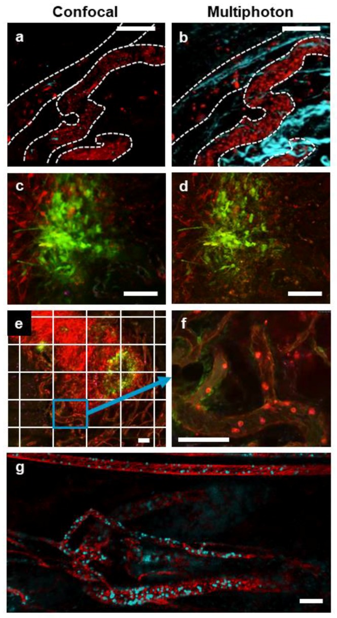
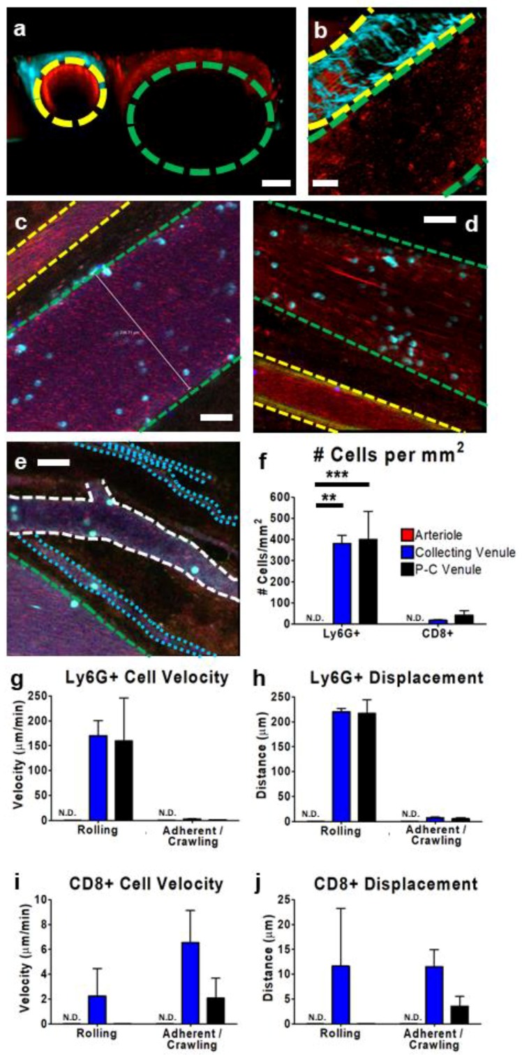
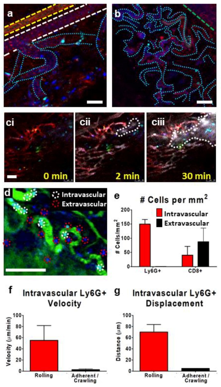
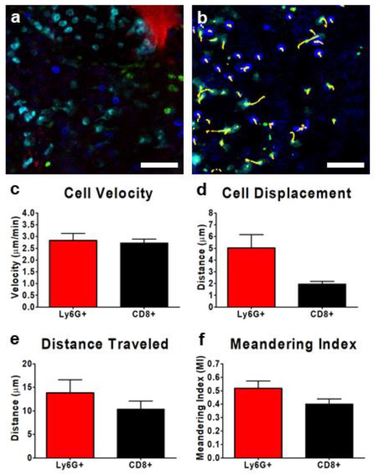
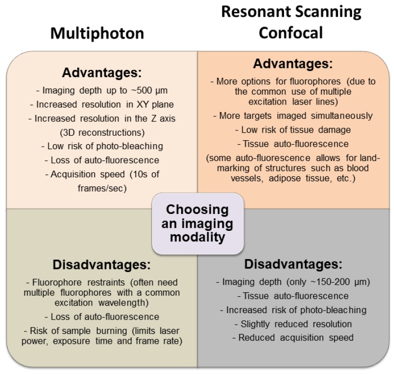
Recent advances in imaging technology have made it possible to track cellular recruitment and behavior within the vasculature of living animals in real-time. Using approaches such as resonant scanning confocal and multiphoton intravital microscopy (IVM), we are now able to observe cells within the intact tumor microenvironment of a mouse. We are able to follow these cells for extended periods of time (hours) and can characterize how specific cell types (T cells, neutrophils, monocytes) interact with the tumor vasculature and cancer cells. This approach provides greater insight into specific cellular behaviors and cell⁻cell interactions than conventional techniques such as histology and flow cytometry. In this report, we describe the surgical preparation of animals to expose the tumor and both resonant scanning confocal and multiphoton imaging approaches used to track leukocyte recruitment, adhesion, and behavior within the tumor microenvironment. We present techniques for the measurement and quantification of leukocyte behavior within the bloodstream and tumor interstitium. The use of IVM to study leukocyte behavior within the tumor microenvironment provides key information not attainable with other approaches, that will help shape the development of better, more effective anticancer drugs and therapeutic approaches.

Keywords: cancer; imaging; intravital; leukocytes; trafficking; vasculature.


Conflict of interest statement

The authors declare no conflict of interest.

Figures

Figure 1
Figure 1
Surgical preparation of subcutaneous and intramuscular tumors for intravital microscopy (IVM) imaging. The mice were injected with tumor cells either subcutaneously on their flank (a) or intramuscularly in the gastrocnemius of the leg (b). After approximately 10 days, the tumors were exposed (aii,bii), tissue movement was surgically stabilized (aiii,biii), and the mouse was inverted and placed onto a heated (37 °C) microscope stage (aiv,biv). An i.v. cannula was inserted into the tail vein to provide anesthetic when necessary throughout the imaging procedure.
Figure 2
Figure 2
IVM of tumor vasculature acquired with resonant scanning confocal or multiphoton imaging modalities. Representative images of a single field of view showing a subcutaneous CT-26 tumor vasculature (red; phycoerythrin (PE)-conjugated anti-CD31 and PE-conjugated anti-CD49b, blood vessels are denoted by a dotted white outline) captured with either confocal (a) or multiphoton (b) imaging (collagen appears blue as a result of second-harmonic generation). In the confocal image (a), some CD8+ cells (blue; eFluor 660-conjugated anti-CD8α) are visible; however, these cells are not seen in the multiphoton image (b) because of a lack of fluorophore stimulation by the single multiphoton excitation wavelength used in this imaging. Confocal (c) and multiphoton (d) imaging of a subcutaneous CT-26 tumor (red) infected with VSV that is transgenic for GFP (VSVΔM51-GFP; green). A stitched, tile scan image (e) of an entire subcutaneous CT-26 tumor (bright red) demonstrating localized VSV infection (green) was captured using resonant scanning confocal imaging. Note each tiled image (outlined by the white grid) was captured as a high-resolution video (f) allowing for later analysis at the cellular level (neutrophils in bright red, endothelium in dim red, i.v.-delivered eFluor 660-labelled VSVΔM51-GFP in blue). High-resolution 4D movies of stitched images (g) of the tumor vasculature of a rhabdomyosarcoma within the gastrocnemius muscle (red; PE-conjugated anti-CD31 and PE-conjugated anti-CD49b), containing neutrophils (cyan; BV421-conjugated anti-Ly6G) captured using multiphoton microscopy. The white scale bar represents 100 µm. Images in (a,c,e,f) were captured using resonant scanning confocal microscopy, whereas images in (b,d,g) were captured using resonant scanning multiphoton microscopy.
Figure 3
Figure 3
Characterization of tumor vasculature and leukocyte behavior within subcutaneous CT-26 tumor vessels. Representative IVM images (ae) show the tumor vasculature. Using multiphoton imaging (a,b), differentiation between arterioles (yellow outline) and collecting venules (green outline) was facilitated by the observation of a collagen sheath (cyan-colored second-harmonic generation) surrounding the arteriole. Vasculature was highlighted by the presence of circulating platelets (red; PE-conjugated anti-CD49b), neutrophils (cyan; BV421-conjugated Ly6G), and CD8+ leukocytes (blue; eFluor 660-conjugated anti-CD8) and was imaged in either cross section (a) or transverse section (b). Using resonant scanning confocal microscopy (ce), arterioles (yellow outline) were apparent as a result of increased autofluorescence (green) and collecting venules (green outline) were seen as parallel unbranching structures. In contrast, post-capillary venules (P-C venules) were observed as narrower, branching vessels (white outline), and tumor microcirculation/capillaries as very narrow (1–2 cell diameter) vessels (cyan outline) that followed a more convoluted path. Quantification of neutrophil (Ly6G+) and cytotoxic T cell (CD8+) interactions (cells present for ≥3 min) within arterioles (red), collecting venules (blue), and post-capillary venules (black) (f). Cell velocity (g,i) and displacement (h,j) of rolling and adherent and crawling neutrophils (g,h) and CD8+ T cells (i,j), as measured over a 10 min imaging period in a subcutaneous CT-26 tumour; n = 3 animals. Data displayed as the mean ± SEM. Total cell counts normalized for the area of each image occupied by a given vessel type. The white scale bar represents 50 µm. Statistical significance was determined using ANOVA; ** = p < 0.01; *** = p < 0.001; N.D. = not detected. Images in (ce) were capture using resonant-scanning confocal microscopy, whereas images in (a,b) were captured using resonant scanning multiphoton microscopy.
Figure 4
Figure 4
Characterization of subcutaneous CT-26 tumor microvasculature and leukocyte behavior. Representative IVM images (ac) show the tumor microvasculature. The vasculature is highlighted by the presence of circulating platelets (red; PE-conjugated anti-CD49b), neutrophils (cyan; BV421-conjugated Ly6G), and CD8+ leukocytes (blue; eFluor 660-conjugated anti-CD8) (a,b). Using resonant scanning confocal microscopy, arterioles (yellow outline, a), veins (green outline, b) were seen as parallel unbranching structures. Venules were seen as narrower than veins and appeared as branching vessels (white outline, a), whereas capillaries/tumor microvasculature appeared as very narrow, convoluted vessels (cyan outline, a,b). Intravenous administration of fluorescent nanoparticles (Q-tracker; grey) identified areas of vascular leakage and accumulation of dye in the tissue interstitium (outlined in dotted white line) (ciciii). The introduction of FITC-conjugated albumin (green) allowed for an easy determination of intravascular (red circles) or extravascular (white circles) leukocytes (d). Quantification of intravascular (red) or extravascular (black) neutrophils (Ly6G+) and cytotoxic T cells (CD8+) (cells present for ≥3 min) within the tumor microvasculature (e). Velocity (f) and displacement (g) of rolling and adherent and crawling neutrophils as measured over a 10 min imaging period in a subcutaneous CT-26 tumor; n = 3 animals. Data displayed as the mean ± SEM. Total cell counts normalized for the area of each region of interest within each image (i.e., vessel vs. extravascular tissue). The white scale bar represents 50 µm. Images in (ad) were captured using resonant scanning confocal microscopy.
Figure 5
Figure 5
Characterization of interstitial leukocyte behavior within the TME of a subcutaneous CD-26 tumor. Representative resonant scanning confocal images of the tumor interstitium (a,b); tumor cells (red), neutrophils (cyan; BV421-conjugated Ly6G), and CD8+ leukocytes (blue; eFluor 660-conjugated anti-CD8) (a,b). Cell movement was tracked over a 10 min imaging window (yellow tracks) for at least three separate fields of view and quantified (b). Leukocyte velocity (c), displacement from initial starting point (d), distance travelled (e), and meandering index (f) were measured for each of the neutrophils (red) and CD8+ T cells (black); n = 3 animals. Between 8 and 50 Ly6G+ cells were traced in each field of view, whereas only 0–4 CD8+ cells were visualized per field of view. Data displayed as the mean ± SEM. The white scale bar represents 50 µm. Images in (a,b) were captured using resonant scanning confocal microscopy.
Figure 6
Figure 6
Comparing resonant scanning confocal versus multiphoton IVM imaging for studying cell recruitment and behavior in the tumor microenvironment (TME). The advantages and disadvantages of each imaging modality are listed to provide a basis for choosing which technique would best suit a specific experiment.

Similar articles

Cited by

References

    1. Cohnheim J. Untersuchungen über die Embolischen Process. August Hirschwald; Berlin, Germany: 1872.
    1. Waller A. Microscopic observations on the perforation of the capillaries by the corpuscles of the blood, and on the origin of mucus and pus-globules. Lond. Edinb. Dublin Philos. Mag. 1846;29:397–405. doi: 10.1080/14786444608645527. - DOI
    1. Hall M. A Critical and Experimental Essay on the Circulation of the Blood. R.B. Seeley and W. Burnside; London, UK: 1831.
    1. Johnston B., Burns A.R., Suematsu M., Issekutz T.B., Woodman R.C., Kubes P. Chronic inflammation upregulates chemokine receptors and induces neutrophil migration to monocyte chemoattractant protein-1. J. Clin. Investig. 1999;103:1269–1276. doi: 10.1172/JCI5208. - DOI - PMC - PubMed
    1. Ho M., Hickey M.J., Murray A.G., Andonegui G., Kubes P. Visualization of Plasmodium falciparum-endothelium interactions in human microvasculature: Mimicry of leukocyte recruitment. J. Exp. Med. 2000;192:1205–1211. doi: 10.1084/jem.192.8.1205. - DOI - PMC - PubMed

LinkOut - more resources