. 2018 Jul 3;23(7):1621.
doi: 10.3390/molecules23071621.

Application of Docking Analysis in the Prediction and Biological Evaluation of the Lipoxygenase Inhibitory Action of Thiazolyl Derivatives of Mycophenolic Acid

Affiliations

Application of Docking Analysis in the Prediction and Biological Evaluation of the Lipoxygenase Inhibitory Action of Thiazolyl Derivatives of Mycophenolic Acid

Evangelia Tsolaki et al. Molecules. .

Abstract

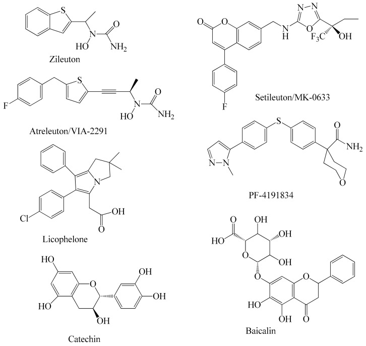
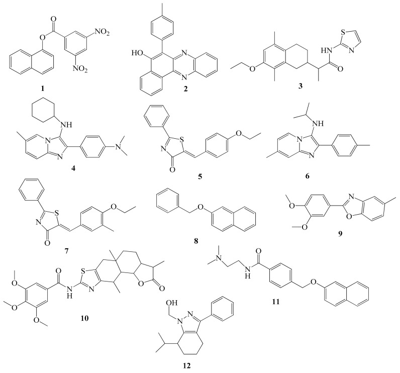
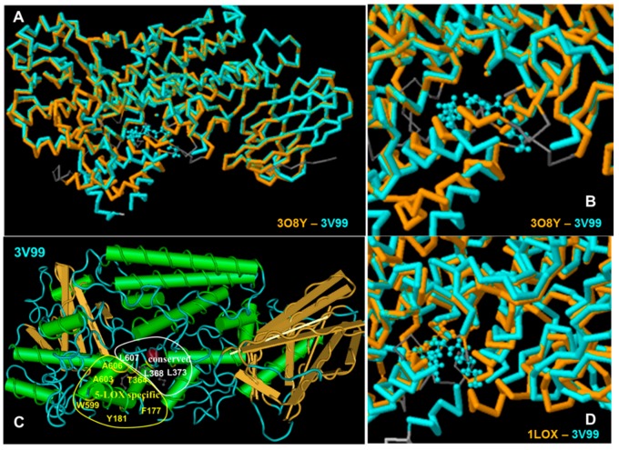
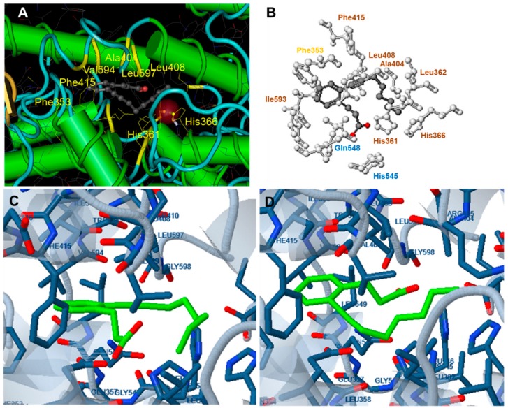
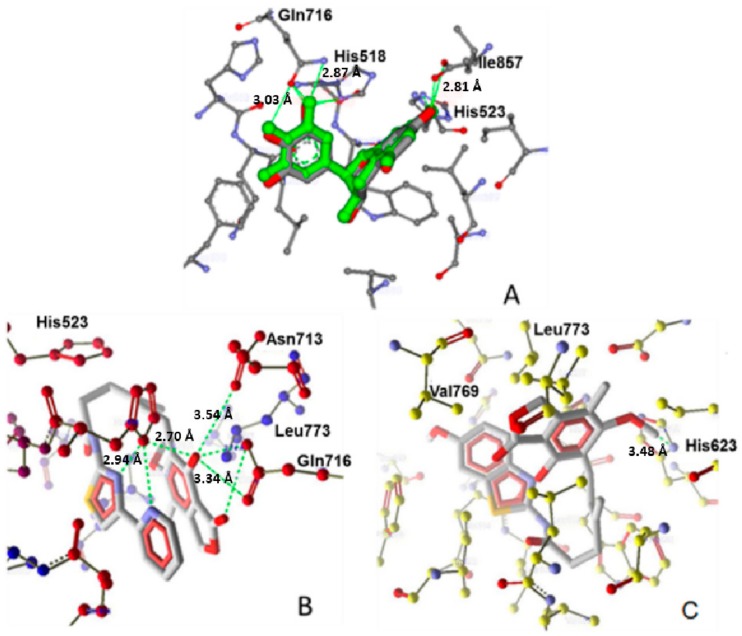
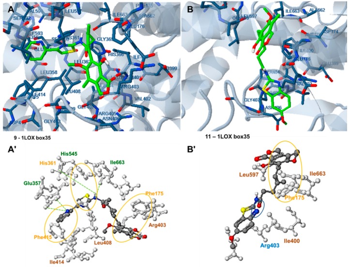
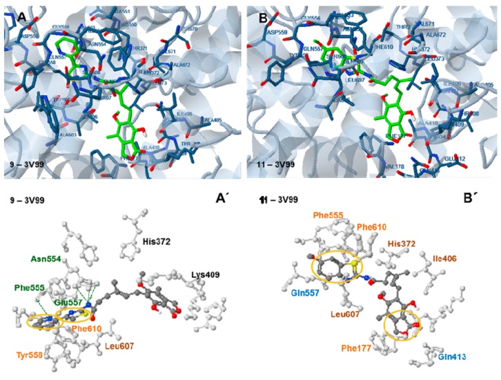
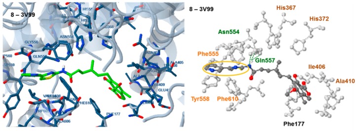
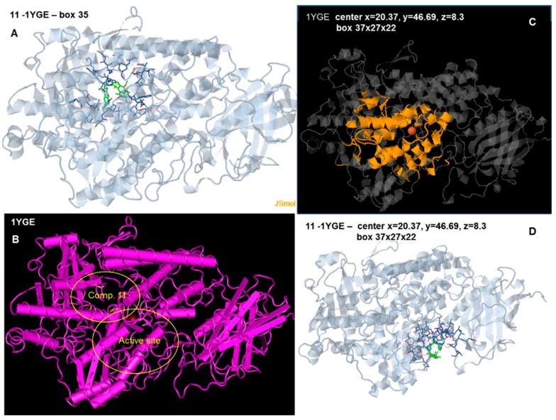
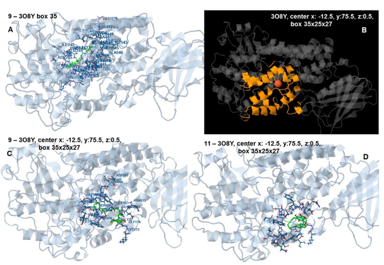
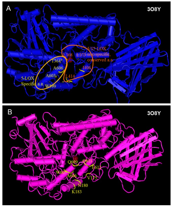
5-LOX inhibition is among the desired characteristics of anti-inflammatory drugs, while 15-LOX has also been considered as a drug target. Similarity in inhibition behavior between soybean LOX-1 and human 5-LOX has been observed and soybean LOX (sLOX) type 1b has been used for the evaluation of LOX inhibition in drug screening for years. After prediction of LOX inhibition by PASS and docking as well as toxicity by PROTOX and ToxPredict sixteen (E)-N-(thiazol-2-yl)-6-(4-hydroxy-6-methoxy-7-methyl-3-oxo-1,3-dihydroisobenzofuran-5-yl)-4-methylhex-4-enamide derivatives with lengths varying from about 15⁻20 Å were evaluated in vitro for LOX inhibitory action using the soybean lipoxygenase sLOX 1b. Docking analysis was performed using soybean LOX L-1 (1YGE), soybean LOX-3 (1JNQ), human 5-LOX (3O8Y and 3V99) and mammalian 15-LOX (1LOX) structures. Different dimensions of target center and docking boxes and a cavity prediction algorithm were used. The compounds exhibited inhibitory action between 2.5 μΜ and 165 μΜ. Substituents with an electronegative atom at two-bond proximity to position 4 of the thiazole led to enhanced activity. Docking results indicated that the LOX structures 1JNQ, 3V99 and 1LOX can effectively be used for estimation of LOX inhibition and amino acid interactions of these compounds.

Keywords: LOX; anti-inflammatory; docking; pharmacophore; thiazoles.


Conflict of interest statement

The authors declare no conflict of interest.

Figures

Figure 1
Figure 1
5-Lipoxygenase inhibitors under clinical development.
Figure 2
Figure 2
Novel 5-lipoxygenase inhibitors identified by computational screening methods.
Figure 3
Figure 3
(A,B) Structure alignment between the human 5-LOX structure 3O8Y (crystalized without substrate or inhibitor) and human 5-LOX structure 3V99 (crystalized with arachidonic acid); (C) Amimno acids of human 5-LOX (structure 3V99), conserved in all LOX enzymes (white region), and 5-LOX specific amino acids (yellow area); (D) Structure alignment between the rat 15-LOX, structure 1LOX (crystalized with inhibitor) and human 5-LOX structure 3V99 (crystallized with substrate).
Figure 4
Figure 4
(A) The reference compound 3-(2-oct-1-ynylphenyl) propanoic acid (OPPA) in complex with 1LOX; (B,D) Docking of OPPA to 1LOX (target box 35). The orientation of the compound is analogue to the original complex (In B. blue: polar interactions, brown: hydrophobic interactions, yellow: pi interactions). In the complex of 1LOX with OPPA (box 35), the carboxylic group of the ligand participates in polar interactions with Gln548 and His545. Hydrophobic interactions with His361, His366, Leu362 Ala404, Leu408 and Phe415 are observed while pi-pi interactions between Phe353 and carbon atoms of the phenyl ring of the ligand also take place; (C) Docking of OPPA to 1LOX (target box 30). The compound occupies an opposite orientation to than in the original complex.
Scheme 1
Scheme 1
Synthesis of the title compounds. R1 and R2 are shown in Table 1.
Figure 5
Figure 5
(A) Alignment and molecular interactions of co-crystallized epigalocatechin and redocked epigalocatechin in the LOX binding site PDB ID 1JNQ; (B) Interactions of the most active compound 9 with LOX protein PDB ID 1JNQ; (C) Molecular interactions of the least active compound 11 with LOX protein PDB ID 1JNQ. Hydrogen bonds are shown in green.
Figure 6
Figure 6
Docking analysis of the most active (IC50 2.5 µM) compound 9, (A,A′) and the less active, (IC50 156.25 µM) compound 11 (B,B′) to the active site of the rat 15-LOX structure PDB ID: 1LOX (target box 35). Green: hydrogen bond interactions, Blue: polar interactions, yellow: pi interactions, brown: hydrophobic interactions.
Figure 7
Figure 7
Docking analysis of the most active (IC50 2.5 µM) compound, 9, (A,A′) and the less active, (IC50 156.25 µM) compound 11 (B,B′) with the active site of the human 5-LOX structure PDB ID: 3V99 (target box 30). Green: hydrogen bond interactions, blue: polar interactions, yellow: pi interactions, brown: hydrophobic interactions.
Figure 8
Figure 8
Docking analysis of compound, 8, with the active site of the human 5-LOX structure PDB ID: 3V99 (target box 30). Green: hydrogen bond interactions, yellow: pi interactions, brown: hydrophobic interactions.
Figure 9
Figure 9
(A,B) Docking of compound 11 to the soybean LOX-1 structure 1YGE, target center: x = 26.37 Å, y = 42.69 Å, z = 12.36 Å, target box 35 × 35 × 35Å. The target box enables the compound to dock preferentially at the upper left corner, behind the active site. The interacting amino acids are marked in yellow at Figure 7B; (C) Structure 1YGE with the target box in yellow (center x = 20.37 Å, y = 46.69 Å, z = 8.3 Å, box 37 × 27 × 22 Å); (D) Docking of compound 11 to 1YGE (center x = 20.37 Å, y = 46.69 Å, z = 8.3 Å, box 37 × 27 × 22 Å).
Figure 10
Figure 10
(A) Docking of compound 9 to the human 5-LOX structure 3O8Y, target center: x = 8.59 Å y = 22.65 Å, z = −1.02 Å, target box 35 × 35 × 35 Å. The target box enables the compound to dock preferentially at the upper left corner, behind the active site; (B) Structure 3O8Y with the target box in yellow (center x: −12.5 Å, y: 75.5 Å, z: 0.5 Å, box 35 × 25 × 27 Å); (C) Docking of compound 9 to 3O8Y (center x: −12.5 Å, y: 75.5 Å, z: 0.5 Å, box 35 × 25 × 27 Å); (D) Docking of compound 11 3O8Y (center x: −12.5 Å, y: 75.5 Å, z: 0.5 Å, box 35 × 25 × 27 Å).
Figure 11
Figure 11
(Α) Conserved and 5-LOX specific amino-acids which participate in interactions with substrates and inhibitors at the active site of 3O8Y, indicated in yellow (some of them barely seen because they are hidden inside the cavity); (B) Amino acids interacting with compound 11 in the preferred docking pose to 308Y (center x: −12.5 Å, y: 75.5 Å, z: 0.5 Å, box 35 × 25 × 27). The interacting amino acids are located at the external area of the molecule.

Similar articles

Cited by

References

    1. Hirata F., Axelrod J. Phospholipid methylation and biological signal transmission. Science. 1980;209:1082–1090. doi: 10.1126/science.6157192. - DOI - PubMed
    1. Roberts L.J. Introduction: Lipids as regulators of cell function. Cell. Mol. Life Sci. 2002;59:727–728. doi: 10.1007/s00018-002-8461-3. - DOI
    1. Kuhn H. Lipoxygenases. In: Marks F., Fustenberger G., editors. Prostaglandins, Leukotrienes and Other Eicosanoids. Wiley-VCH; Weinheim, Germany: 1999. p. 109.
    1. Radmark O., Samuelsson B. 5-Lipoxygenase: Mechanisms of regulation. J. Lipid Res. 2009;50:S40–S45. doi: 10.1194/jlr.R800062-JLR200. - DOI - PMC - PubMed
    1. Schneider C., Pratt D.A., Porter N.A., Brash A.R. Control of oxygenation in lipoxygenase and cyclooxygenase catalysis. Chem. Biol. 2007;14:473–488. doi: 10.1016/j.chembiol.2007.04.007. - DOI - PMC - PubMed

MeSH terms

LinkOut - more resources